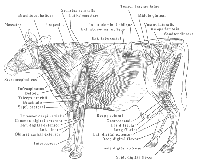
The Superficial Muscles of a Cow ClipArt ETC

MODEL OF A COW'S ANATOMY, THE MUSCLES, FRAGONARD MUSEUM, NATIONAL
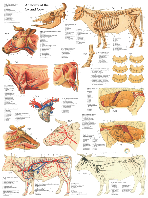
The superficial muscles of a cow are diagramed. Labels: 1, Occipito-Frontalis. 2, Orbicularis Palpaebrarum. 3, Masseter. 5, Sterno-cleido-Mastoid. 6, Trapezius. 7, Latissimus Dorsi. 8, Pectoralis. 9, 10, External and Internal oblique muscles. 11, Opening of the mammary artery and vein (milk vein). 12, Gluteii. 13, Rectus Femoris muscle.

Very Muscled Cow In Green Field by Compuinfoto Green fields, Cow
It is a triangular depression bounded by lumbar transverse processes and the epaxial muscle dorsal to the processes, the last rib, and an oblique muscular thickening of the internal abdominal oblique m. extending from the tuber coxae to the costal arch. (see image below)

Cow muscles Mammals, Anatomy for artists
1, masseter muscle; 2, coronoid process; 3, temporal fossa; arrowheads, temporal line; 4, paracondylar process; 5, occipital condyle; 6-9 cheek teeth (Triadan numbers).. Figure 25-18 Left half of upper and right half of lower jaw of cow. Note the different shapes of the upper and lower cheek teeth and the large diastema (1).

Anatomia geral do touro e da vaca Atlas ilustrado
The muscles of the shoulder include the deltoid muscles, teres major, teres minor, supraspinatus, infraspinatus, subscapularis and coracobrachialis. These muscles provide flexion and stability to the shoulder joint. The elbow joint extensors include the triceps brachii and the tensor fasciae antebrachii.
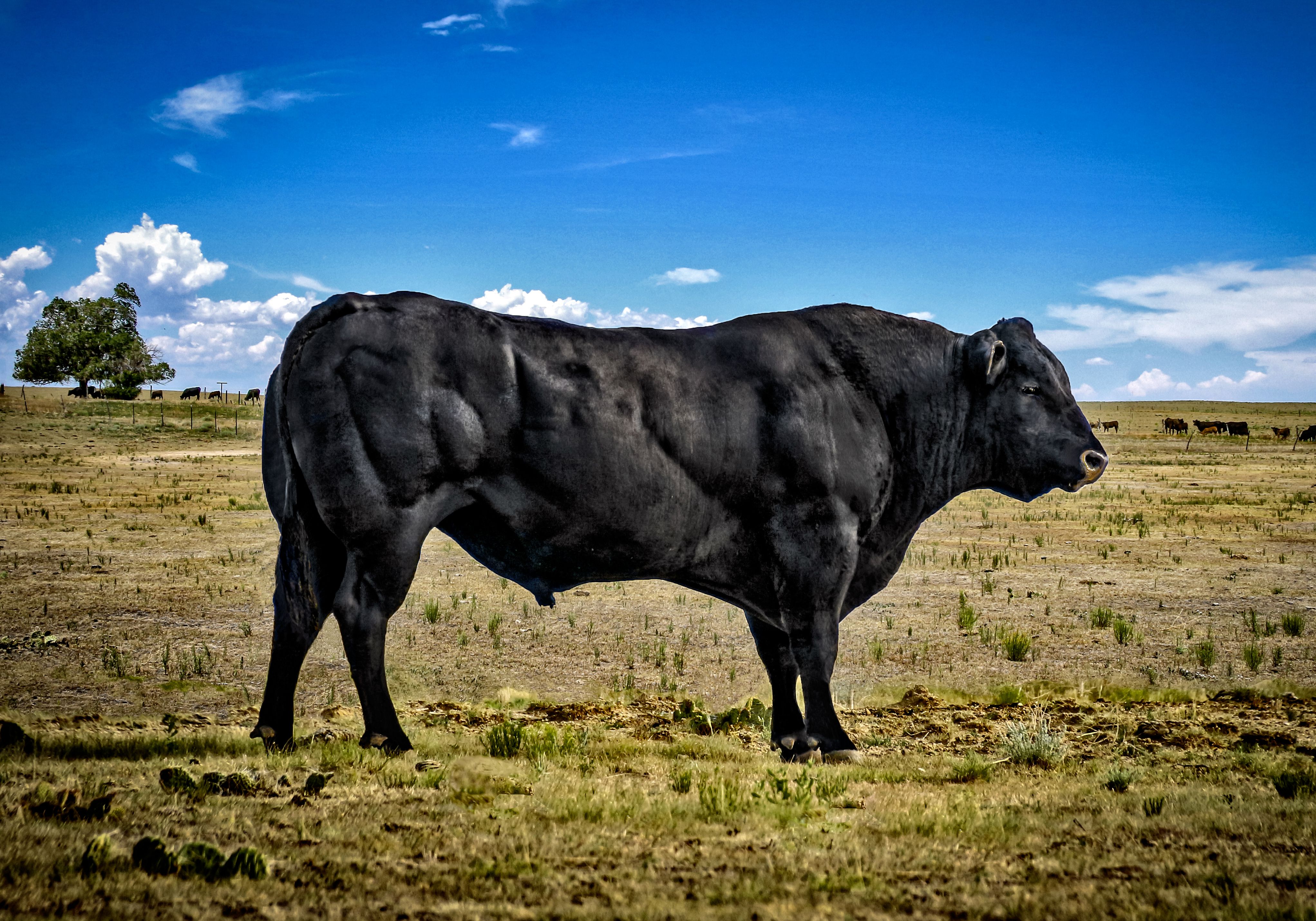
Muscular Cow Pictures All About Cow Photos
From the top of the head and along the top side of the cow, the skeletal system includes the horn cones, cervical vertebrae, dorsal vertebrae, lumber vertebrae, sacrum and hip bone. Along the back side of the cow, points of interest on the cow's skeletal system include: the femur. knee joint. tibia. hock joint.

Bovine Cow Muscle Anatomy Poster in 2021 Muscle anatomy
A baby cow is called a calf. A female calf is sometimes called a heifer calf and a male a bull calf. A heifer is a female that has not had any offspring. The term usually refers to immature females; after giving birth to her first calf, however, a heifer becomes a cow. An adult male is known as a bull.
The Superficial Muscles of a Cow ClipArt ETC
Muscles of thoracic limb of a cow Muscles of the hindlimb of a cow Cow anatomy organs Digestive organs of a cow Cow anatomy stomach Compartments of cow stomach Liver and pancreas of cow anatomy Organs of the respiratory system from a cow Lung anatomy of a cow Heart of a cow Cow hoof anatomy Cow anatomy labeled diagram
musclecows005 Built Report
These are the gastrocnemius and soleus, (the 'calf muscles' in humans). Some of the more fragile edges of this calcaneus are missing, but you can still see the main features. This photo is pretty much a close-up of the photo above, from the bottom end. © Saffron Walden Museum.
Anatomy
Quick overview: there are several muscles in the body of a cow, but I will identify the most used and useful muscles from the head, neck, thorax, abdomen, and limbs. Muscles from the neck and limbs are most important for field practices.
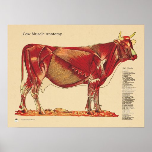
Cow Bovine Veterinary Muscles Anatomy Chart Poster Zazzle
Category: Anatomy This chart shows views of the cow's left lateral view with the dorsal and vertebral regions indicated. In addition, superficial muscles and the cow's veins, deep cervical muscles, major joints, in situ viscera, and udder are also shown.
Cow Ox Anatomy Poster
In some breeds of cows, such as the Jersey, the dewclaws may be more pronounced and may even have small hooves attached. Chest floor. The chest floor of a cow contains several important muscles that are used for movement and support. Elbow. The elbow of a cow can be injured if the animal falls or experiences trauma to the front leg. Fore udder

183 best Anatomie Bovine images on Pinterest Animal anatomy, Cow and Cows
A3.2 Identify and describe the joints, joint angles, joint actions, and muscle groups of the pelvic limb. Joints of the pelvic (hind) limb. Clinical Notes: joint pouches are extensions of the synovial capsule and cavity past joint surface. In more mobile joints these pouches can be more expansive/extensive.
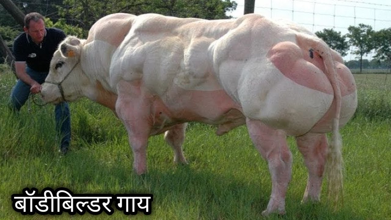
ये है बॉडीबिल्डर गाय, कभी देखी हैं आपने The World's Biggest
25/04/2023 28/10/2022 by Sonnet Poddar The cow leg anatomy consists of bones, muscles, nerves, and vessels. Bones are the hardest and main component of the cow leg structure. Again, the muscles are also essential as most vessels and nerves pass along or within them.
musclecows026 Built Report
1 Pelvic Girdle and Hip 1.1 Bones 1.1.1 Bovine Bone Specifics 2 Joints and Synovial Structures 2.1 Sacroiliac Joint 2.2 Coxafemoral/Hip Joint 3 Musculature 4 Proximal Hindlimb including Stifle and Tarsus 4.1 Bones 4.1.1 Bovine Bone Specifics 4.2 Joints and Synovial Structures 4.3 Musculature 5 Vasculature of the Hindlimb 6 Webinars

Muscle cow by Goutofang1 on DeviantArt
Despite its name, the is located laterally in meat animals. It covers the lateral face of the ilium and appears as the large muscle area in sirloin steaks and chops. The flank and belly of the animal are formed by sheets of muscle and connective tissue.

cow anatomy study, Robin de Jong Anatomy study, Creature design
5 Muscles of the Forelimb 5.1 Extrinsic Musculature 5.2 Intrinsic Musculature 6 Muscles of the Shoulder 6.1 1. Lateral 6.2 2. Medial 6.3 3. Caudal (Flexors) 7 Muscles of the Elbow 7.1 Extensors 7.2 Flexors 8 Muscles of the Carpal and Digital Joints 8.1 Extensors 8.2 Flexors 9 Vasculature of the Forelimb 10 Webinars
