Facial Muscles and Expressions Classic Human Anatomy in Motion The

Human Muscle Anatomy Head
Anatomy Facial muscles Author: Gordana Sendić MD • Reviewer: Jana Vasković MD Last reviewed: November 21, 2023 Reading time: 27 minutes Recommended video: Muscles of facial expression [12:24] Overview of the muscles responsible for facial expression. Facial muscles (Musculi faciales)

Pin on Anatomy for Sculpture The Human Neck
How to remember every muscle in the head and face.Visit https://khub.me/corporis to get Kenhub for 10% offThanks to the @Vlogbrothers scholarship for support.

Muscle diagram, Muscles of the face, Muscle anatomy
Key Terms. depressor labii inferioris: An analogous muscle that lowers the bottom lipEndFragment; Buccinator: This muscle is located between the upper and lower jaws in the cheek, deep to the other muscles of the face.; zygomatic: This muscle controls the cheeks to create smiles and frowns.; Procerus: The most superior of all facial muscles.; depressor anguli oris: This muscle is opposite to.

Pin on Stacy PAIN TOS
The facial muscles can broadly be categorised into three groups - orbital, nasal and oral. In this article, we shall look at the anatomy of the muscles of facial expression - their attachments, actions and clinical relevance. Fig 1 - Innervation to the muscles of facial expression via the facial nerve (CN VII) Orbital Group
Face And Neck Muscle Diagram / Facial Muscles Images Stock Photos
Face muscle anatomy Test your information with labeled diagrams Practice test Interactive headmost muscles quizzes Sources + Show choose Face string anatomy

Jeff Searle Head muscles, Human body anatomy, Facial muscles anatomy
Learn and practice the facial muscles more effectively using our facial muscles quizzes and labeled diagrams.

fac06_15FigureL.jpg (1360×1050) Neck muscle anatomy, Facial muscles
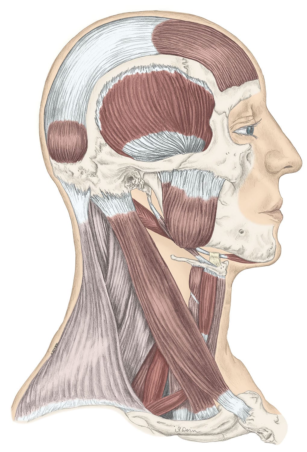
The muscles of the face overlap and crisscross over each other, creating a mask of muscle over the skull and jawbone. They attach to various parts of the skull and other muscles, allowing for a.

Anatomy of the facial muscles. Reprinted under Creative Commons
Test your knowledge with labeled diagrams Now that you've learned about the muscles of the face, it's time to test your memory and understanding. The best way to do this at the beginning of your revision is by using labeled diagrams.
Labeled Chart Of The Facial Muscles Photograph by Hank Grebe Fine Art
Muscles and Tendons Information Center Reviewed By: Pramod Kerkar, M.D., FFARCSI, DA There are about 20 flat skeletal muscles that construct the facial structure. All of these muscles have different functions in the face. Innervated by the cranial nerve, which is the facial nerve, the muscles control all of our facial expressions.

Head Muscles Diagram Face muscles anatomy, Anatomy
The human face is the most anterior portion of the human head. It refers to the area that extends from the superior margin of the forehead to the chin, and from one ear to another. The basic shape of the human face is determined by the underlying facial skeleton (i.e. viscerocranium ), the facial muscles and the amount of subcutaneous tissue.

Facial Muscles and Expressions Classic Human Anatomy in Motion The
Go to: Structure and Function The anatomy of the face can divide into three main regions: upper face, middle face, and lower face. The entire face is covered by skin superficially, while the deep anatomy contains muscles, fat pads, nerves, vessels, and bones. Upper Face

Human Muscles Diagrams Labeled 2019 101 Diagrams
Facial Muscles Facial muscles work together to control the parts of your face. They are essential to chewing and making facial expressions. If you experience weakness or paralysis in your face muscles, seek medical attention. Although facial palsy can be a sign of a temporary, curable condition, it may also indicate a serious medical problem.

Muscles of Face Anatomy Flashcards Anatomic.us Muscles of Face
Muscles of Facial Expression Blood Supply: External Carotid Artery Motor Innervation: Facial Nerve (Vll) Sensory Innervation: Trigeminal Nerve (V) Frontalis (worry muscle): Actions: Raises eyebrows, furrows brow Innervation: Facial Nerve (Vll) Origin: from galea aponeurotica Insertion: to skin above the eyebrows

Vintage Human Anatomy Muscles (Face, Head, Neck) Poster
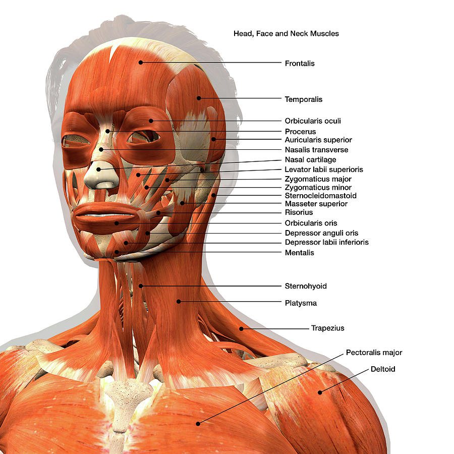
Interactive diagram of the muscles of facial expression, including the frontalis, temporalis, orbicularis oculi, zygomaticus major, zygomaticus minor, nasalis, levator labii superioris, masseter, levator anguli oris, buccinator, obicularis oris and others.

Axial Muscles of the Head, Neck, and Back · Anatomy and Physiology
The neck muscles, including the sternocleidomastoid and the trapezius, are responsible for the gross motor movement in the muscular system of the head and neck. They move the head in every direction, pulling the skull and jaw towards the shoulders, spine, and scapula. Working in pairs on the left and right sides of the body, these muscles.

Pin on Botox
We've just released a collection of 500+ OSCE Stations! 🙌 https://geekymedics.com/osce-stations/ This video provides an overview of the muscles of facial expression using high-quality 3D.
