15 Microscope Parts A Guide on their Location and Function
1.5 Microscopy Biology LibreTexts
This simple worksheet pairs with a lesson on the light microscope, where beginning biology students learn the parts of the light microscope and the steps needed to focus a slide under high power.. The labeling worksheet could be used as a quiz or as part of direct instruction. Students label the microscope as you go over what each part is used for.
15 Microscope Parts A Guide on their Location and Function
Magnification is a measure of how much larger a microscope (or set of lenses within a microscope) causes an object to appear. For instance, the light microscopes typically used in high schools and colleges magnify up to about 400 times actual size. So, something that was 1 mm wide in real life would be 400 mm wide in the microscope image.

301 Moved Permanently
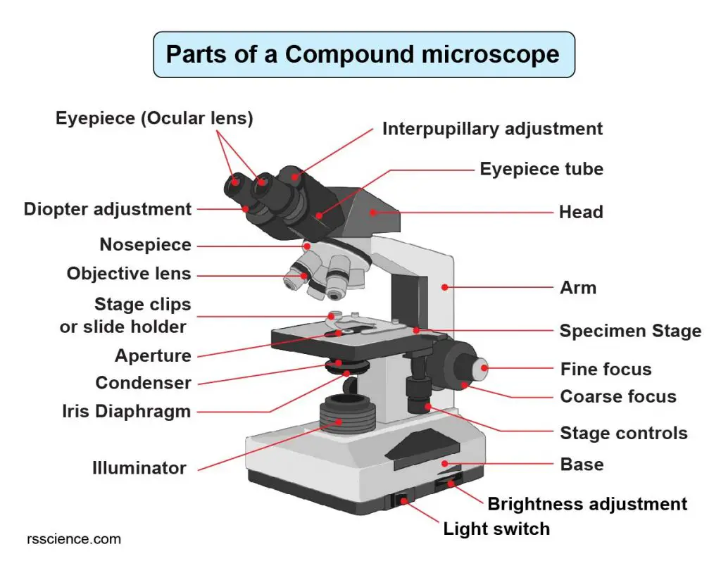
Parts of a Compound Microscope Each part of the compound microscope serves its own unique function, with each being important to the function of the scope as a whole. The individual parts of a compound microscope can vary heavily depending on the configuration & applications that the scope is being used for. Common compound microscope parts include: Compound Microscope Definitions for.
Clipart microscope parts labeled WikiClipArt
All microscopes share features in common. In this interactive, you can label the different parts of a microscope. Use this with the Microscope parts activity to help students identify and label the main parts of a microscope and then describe their functions.. Drag and drop the text labels onto the microscope diagram. If you want to redo an answer, click on the box and the answer will go back.
Parts of a Microscope SmartSchool Systems
There are 1000 millimeters (mm) in one meter. 1 mm = 10 -3 meter. There are 1000 micrometers (microns, or µm) in one millimeter. 1 µm = 10 -6 meter. There are 1000 nanometers in one micrometer. 1 nm = 10 -9 meter. Figure 1: Resolving Power of Microscopes. The microscope is one of the microbiologist's greatest tools.
The Parts of a Compound Microscope and How To Handle Them Correctly Human Anatomy and
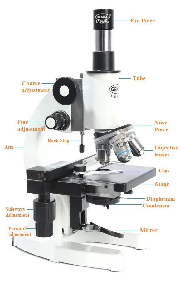
Eyepiece lens magnifies the image of the specimen. This part is also known as ocular. Most school microscopes have an eyepiece with 10X magnification. 2. Eyepiece Tube or Body Tube. The tube hold the eyepiece. 3. Nosepiece. Nosepiece holds the objective lenses and is sometimes called a revolving turret.

Parts of a microscope with functions and labeled diagram
No matter what you love, you'll find it here. Search Microscope spares and more. We've got your back with eBay money-back guarantee. Enjoy Microscope spares you can trust.
Parts Of Microscope
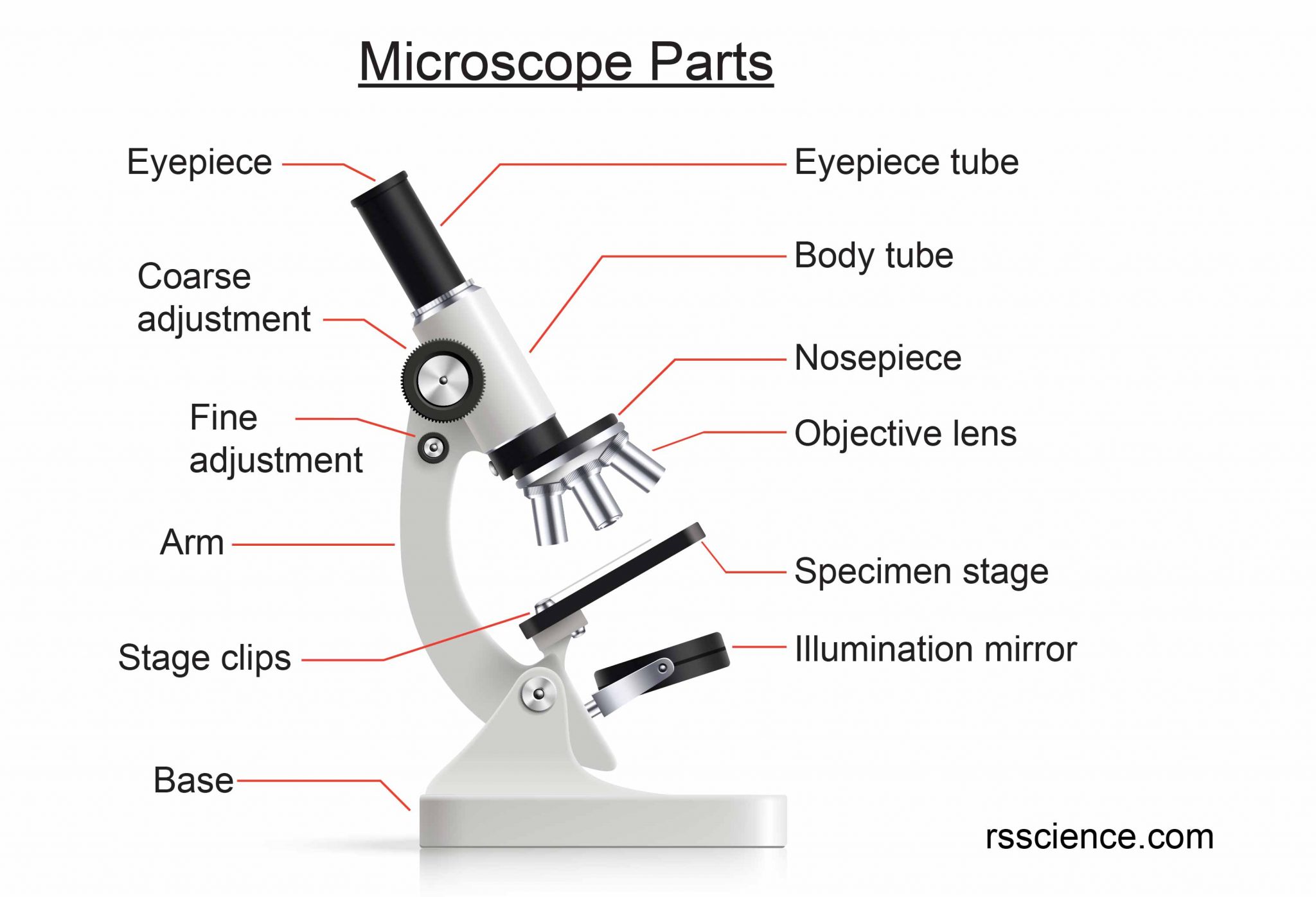
Tube: Connects the eyepiece to the objective lenses. Arm: Supports the tube and connects it to the base. Base: The bottom of the microscope, used for support. Illuminator: A steady light source (110 volts) used in place of a mirror. If your microscope has a mirror, it is used to reflect light from an external light source up through the bottom.
Compound Microscope Parts Labeled Diagram and their Functions (2023)
Parts of Simple Microscope (Labeled Pictures) It is a magnifying glass with a double convex lens and has a distinct short focal length. It magnifies the object being studied through angular magnification. Below are the vital and complete parts of a simple microscope. Eyepiece/Ocular.

Microscope diagram Tom Butler Technical Drawing and Illustration Projects Pinterest
Blank microscope to label parts. This page titled 1.5: Microscopy is shared under a CC BY 4.0 license and was authored, remixed, and/or curated by Orange County Biotechnology Education Collaborative ( ASCCC Open Educational Resources Initiative ) .

Parts of a Microscope and their function
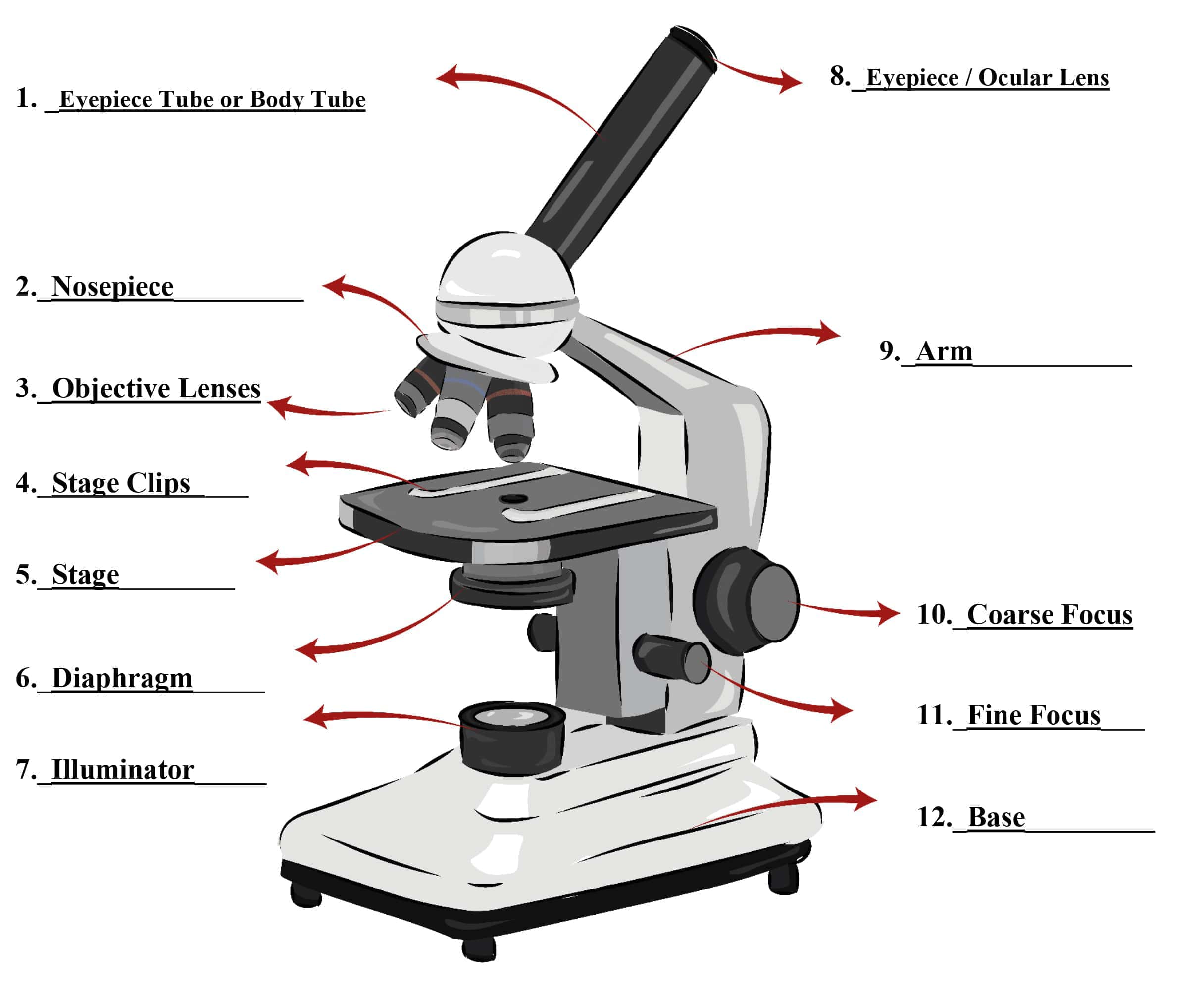
Microscope Parts Labeled: Parts of A Microscope. 1. Eyepiece Lens and Eyepiece Tube. This part is the one on top where users view or look through the device; the eyepiece tube is the tube that connects the ocular lens to the device itself. 2. Objective Lens.
Proper Use & Care of Microscopes Clinician's Brief
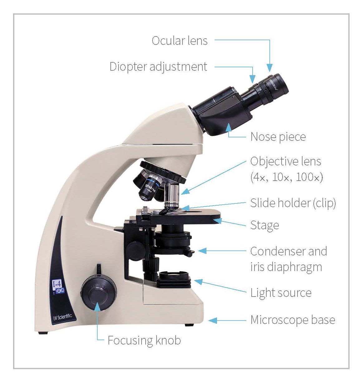
The optical microscope often referred to as the light microscope, is a type of microscope that uses visible light and a system of lenses to magnify images of small subjects. There are two basic types of optical microscopes: Simple microscopes. Compound microscopes. The term "compound" in compound microscopes refers to the microscope having.

Parts of a Compound Microscope Labeled (with diagrams) Medical Pictures and Images (2023
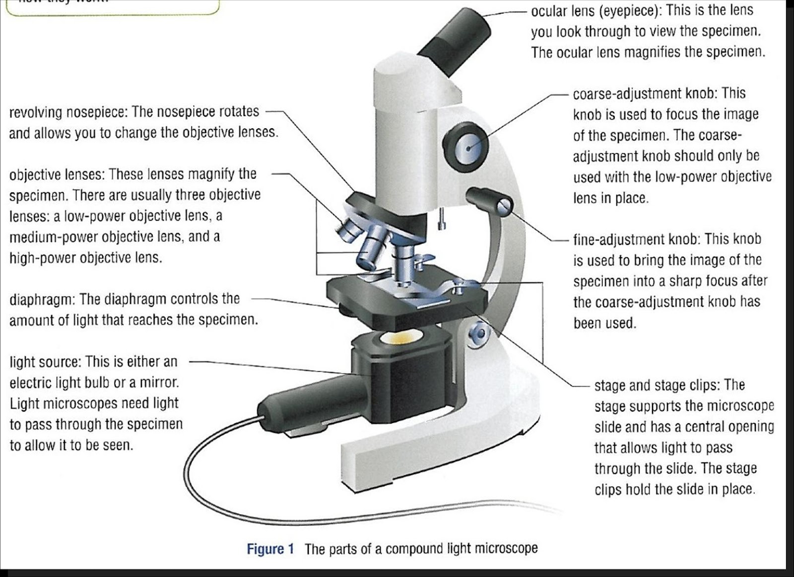
Ocular Lens (eye-piece) Ocular lens of a microscope. It is located at the top of the microscope, and the ocular lens or eyepiece lens is used to look through the specimen. It also magnifies the image formed by the objective lens, usually ten times (10x) or 15 times (15x). Usually, a microscope has an eyepiece of 10x magnification power.
13 Tips You Should Know about Taking Care of Your Microscope Rs' Science
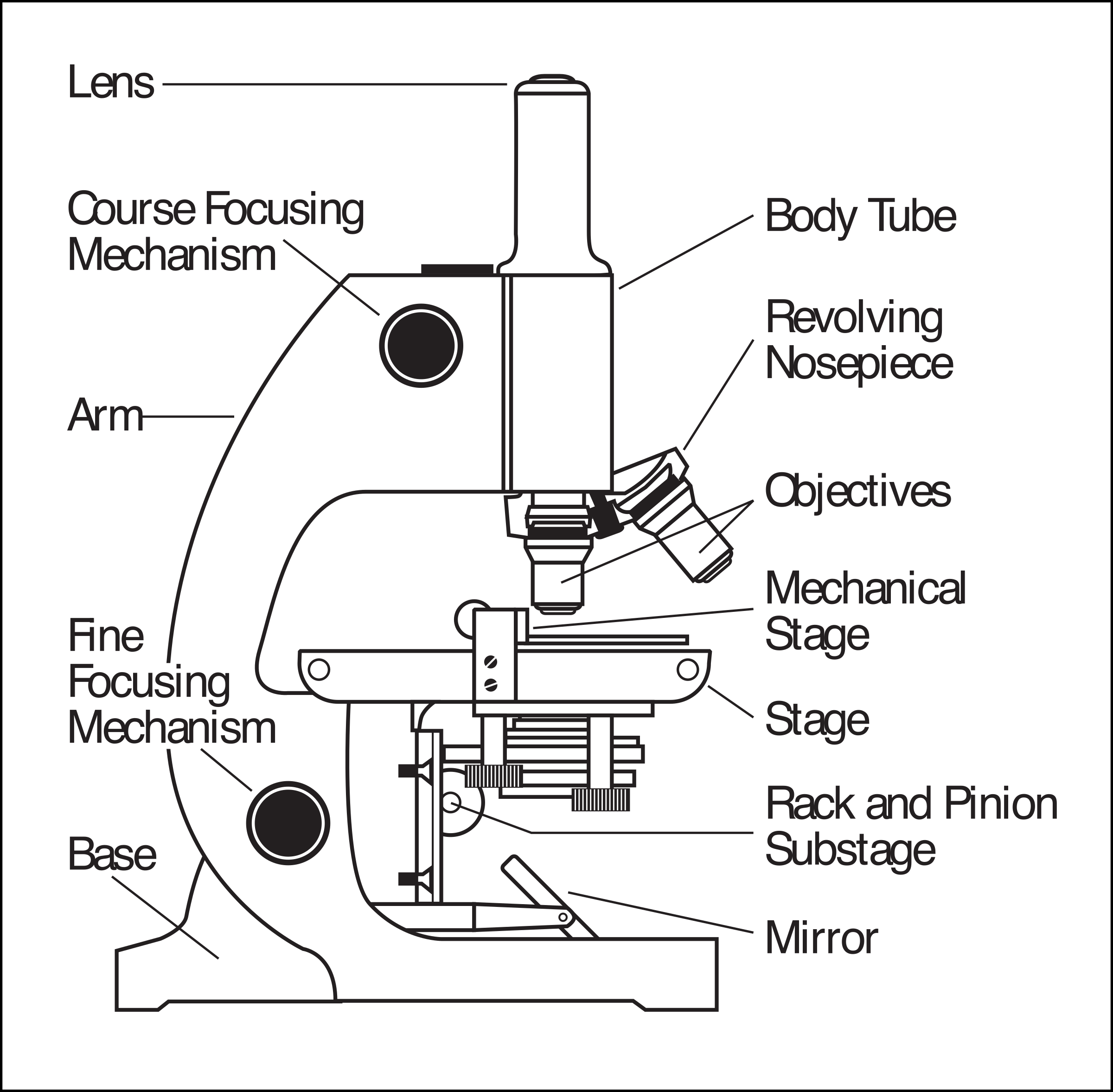
Diopter Adjustment: Useful as a means to change focus on one eyepiece so as to correct for any difference in vision between your two eyes. Body tube (Head): The body tube connects the eyepiece to the objective lenses. Arm: The arm connects the body tube to the base of the microscope. Coarse adjustment: Brings the specimen into general focus.

Simple Microscope Definition, Principle, Magnification, Parts, Applications
B. NOSEPIECE microscope when carried Holds the HIGH- and LOW- power objective LENSES; can be rotated to change MAGNIFICATION. Power = 10 x 4 = 40 Power = 10 x 10 = 100 Power = 10 x 40 = 400 What happens as the power of magnification increases?

Parts of a Microscope The Comprehensive Guide Microscope and Laboratory Equipment Reviews
Labeling the Parts of the Microscope. This activity has been designed for use in homes and schools. Each microscope layout (both blank and the version with answers) are available as PDF downloads. You can view a more in-depth review of each part of the microscope here.
