Orbits and eyes anatomical illustrations eAnatomy

Choroid Function In Eye Map Of Body
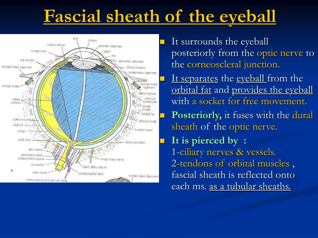
Fascial Sheath of the Eyeball Connective Tissue Fascial Sheath of the Eyeball Vagina bulbi Structure Anatomical Relations Function List of Clinical Correlates Structure The fascial sheath of the eyeball surrounds the eye, extending from the optic nerve to the corneoscleral junction.

Orbit und Auge anatomische Abbildungen
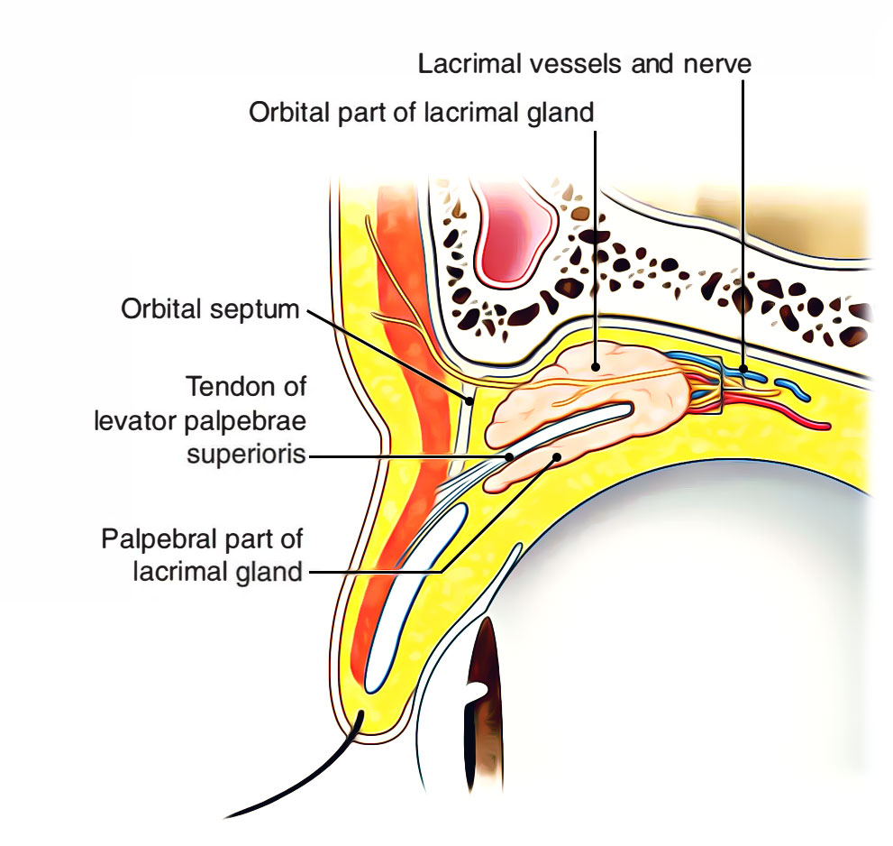
Eyelid anatomy Lacrimal gland Eye muscles Eyeball Outer layer Middle layer Inner layer Blood supply of the eye Nerves of the eye Sources + Show all Bones of the orbit The bony orbit is made out of seven bones, which include the maxilla, zygomatic bone, frontal bone, ethmoid bone, lacrimal bone, sphenoid bone and palatine bone.
PPT Coats of the eyeball PowerPoint Presentation, free download ID4368785
Describe the fascia of eyeball and orbit.What is Tenon's capsule?What is ligament of lockwood's?What is periorbita?Formation of check ligaments?location and.

Orbits and eyes anatomical illustrations eAnatomy
The fascial sheath of the eyeball—also called the Tenon capsule—is a fibroelastic layer that surrounds the entire scleral portion of the globe. It delimits the episcleral space or sub-Tenon space, a potential space with no actual volume, although fluid can be injected into it. Some experts assimilate it into the articular capsule of the globe.
Eyeball Structure and function Kenhub
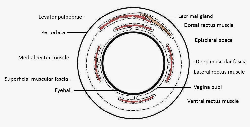
The eyeball and all of the extraocular muscles are enveloped by a muscular fascia, derived from the fascial sheath of the eyeball (Tenon's capsule). This is an important clinical point due to the fact that the subtendinous space is a common location for the injection of local anesthetics during various surgical procedures in this region.
Easy Notes On 【Orbit】Learn in Just 4 Minutes!
The eyes are responsible for detecting visible light, which ranges from 400 to 700 nanometers in wavelength. Objects can absorb and reflect different wavelengths of light. An object appears white if it reflects all wavelengths of light, and it appears black if it absorbs all wavelengths of light.
Suspensory and Test Ligaments of the Eye Earth's Lab
Tenon's capsule is a fascial sheath that encloses the eye, separating the sclera from the conjunctiva anteriorly and the orbital fat posteriorly. There is a potential space between Tenon's layer and the underlying sclera, although the two are firmly adherent to each other approximately 1.5 mm behind the limbus (the junction between sclera and cornea) and at the entry site of the optic.

Fascial sheath of eye ball Tenon capsule capsule of Tenon Tenon's capsule YouTube
Fascial sheath of eyeball (Tenon) : Orbital cavity Extraocular muscles; Extrinsic muscles of eyeball Lacrimal apparatus : Orbital septum Eyeball/Eye : General Anatomy Lens (Eye) : Histology Iris : Anterior view Iridocorneal angle : Gonioscopy Corona ciliaris : Posterior view Retina : Histology
Eye Opener Anatomy Ocular Adnexa
External to the sclera, the eyeball is enveloped by a thin fascial sheath (so-called Tenon's capsule) that extends from the optic nerve to the sclerocorneal junction (fig. 46-3). The sheath separates the globe from the orbital fat and acts as a socket in which the eye moves as in a ball-and-socket joint. It blends with the sheaths of the.

How to perform a lateral canthotomy EyeGuru
Overview Fascial sheath (Tenon's capsule) Function Fibrous layer Sclera Cornea Vascular layer (Uvea) Choroid Ciliary body Iris Nervous layer (Retina) Neural retina Retinal pigment epithelium (RPE) Blood supply Refractive media of the eyeball Lens Vitreous body Aqueous humor Clinical conditions
Suspensory and Test Ligaments of the Eye Earth's Lab
fas·ci·al sheath of eye·ball. A condensation of connective tissue on the outer aspect of the sclera from which it is separated by a narrow cleftlike episcleral space; the sheath is attached to the sclera near the sclerocorneal junction and blends with the fascia of the extraocular muscles. Synonym (s): Tenon capsule.
Orbits and eyes anatomical illustrations eAnatomy
The posterior myofascial chain (PMC) or superficial back line encompasses a series of muscles interlinked by the deep fascia, extending from the foot to the fascial sheath of the eyeball. The deep cervical fascia of the neck, the epicranial aponeurosis of the head, and the fascial sheath of eyeball,.

04. Eye Movement Training Speedy Eyes
Tenon's capsule ( / təˈnoʊn / ), also known as the Tenon capsule, fascial sheath of the eyeball ( Latin: vagina bulbi) or the fascia bulbi, is a thin membrane which envelops the eyeball from the optic nerve to the corneal limbus, separating it from the orbital fat and forming a socket in which it moves.
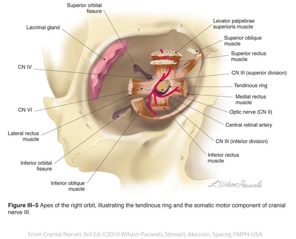
The orbit Extraocular muscle annulus RANZCRPart1 Wiki Fandom
Summary This chapter contains sections titled: Fascial Sheath of the Eyeball (Fascia Bulbi , Tenon's Capsule) The Eyeball Refractive Media of the Eye Clinical Problems Answers to Clinical Problems. Skip to Article Content; Skip to Article Information; Search within. Search term. Advanced.

Orbits and eyes anatomical illustrations eAnatomy
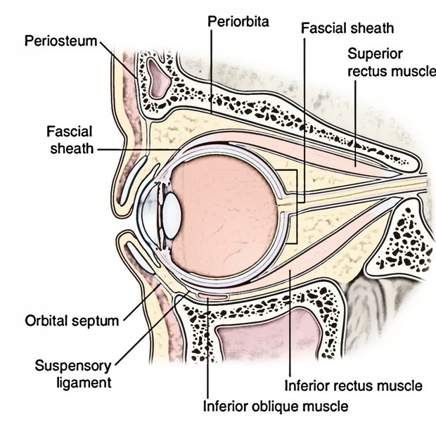
The inserting muscle fibers pierce the fascial sheath of the eyeball (Tenon's capsule), which in turn reflects back and creates a thin fascial sleeve around the muscle's tendon. This sleeve gives off an expansion called the medial check ligament that connects the medial rectus muscle with the medial wall of orbit.

Fasciae of Orbit and Eyeball Anatomy Horizontal section, Medial palpebral ligament, Nasal cavity
Tenon's capsule ( / təˈnoʊn / ), also known as the Tenon capsule, fascial sheath of the eyeball ( Latin: vagina bulbi) or the fascia bulbi, is a thin membrane which envelops the eyeball from the optic nerve to the corneal limbus, separating it from the orbital fat and forming a socket in which it moves. The right eye in sagittal section.
