Anatomy of Uvea

Cilioretinal arteries (CAs) deriving from the short posterior ciliary... Download Scientific
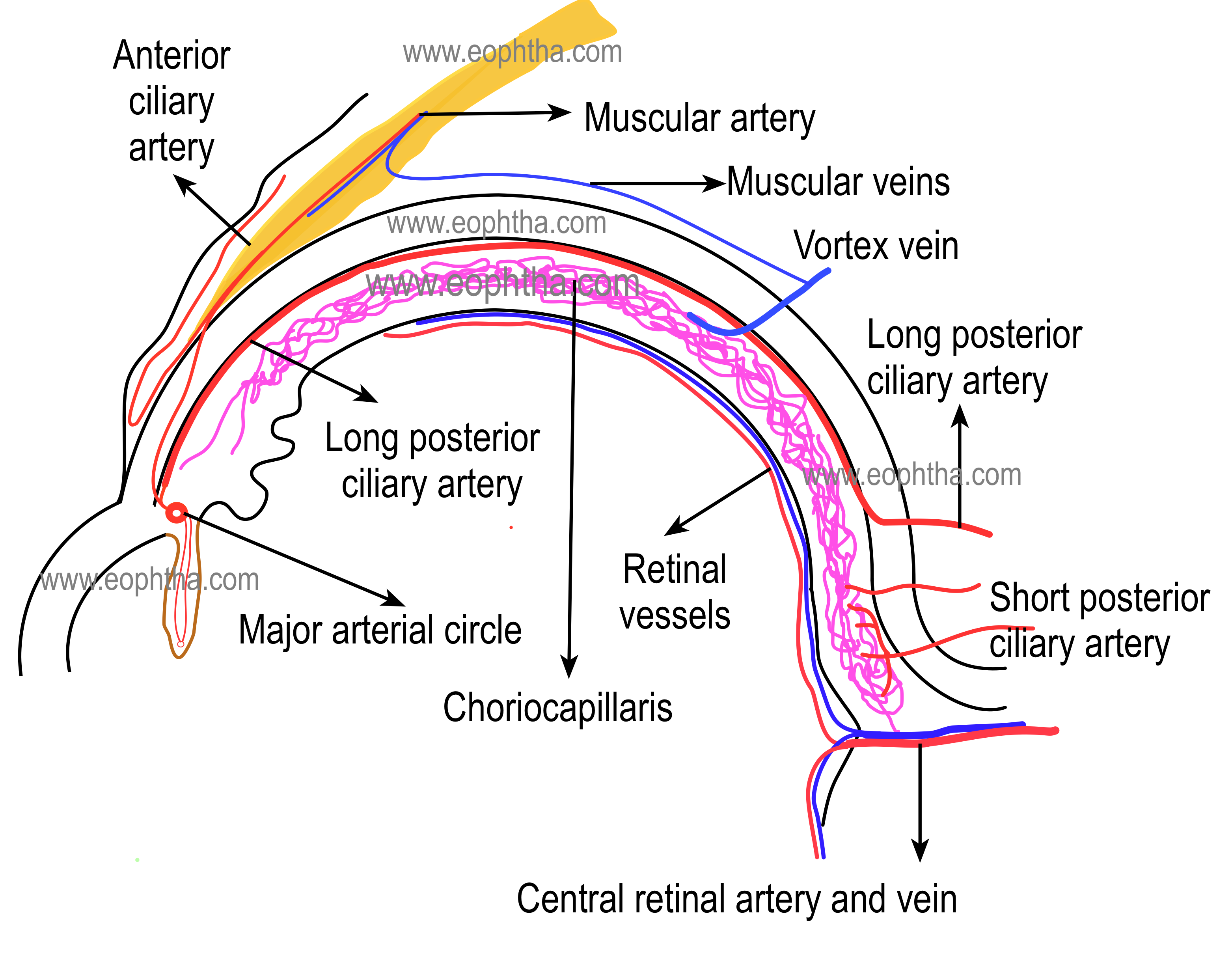
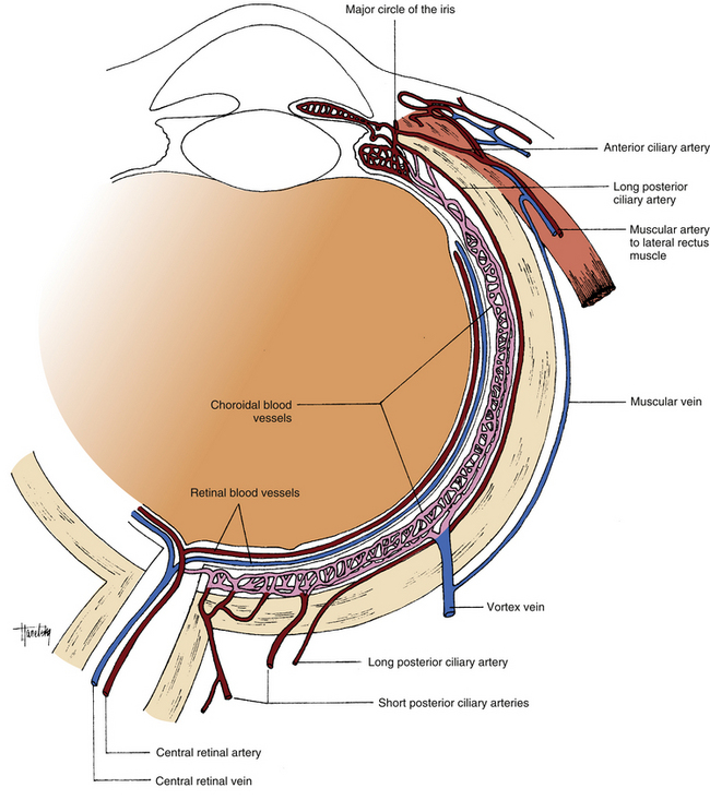
The posterior ciliary artery (PCA) circulation is the main source of blood supply to the optic nerve head (ONH), and it also supplies the choroid up to the equator, the retinal pigment epithelium (RPE), the outer 130 μm of retina (and, when a cilioretinal artery is present, the entire thickness of the retina in that region), and the medial and lateral segments of the ciliary body and iris.
Anatomy of Uvea
(a) Cross sectional B scan flattened around the COI that shows entry of short posterior ciliary artery (SPCA) (arrow) (1:1 pixel); (b) En-face scan obtained at the chorio-scleral junction showing.
Optician Online CPD Archive
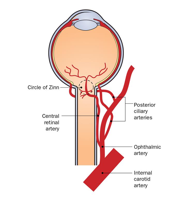
The short posterior ciliary arteries are a number of branches of the ophthalmic artery. They pass forward with the optic nerve to reach the eyeball, piercing the sclera around the entry of the optic nerve into the eyeball. Anatomy
Short Posterior Ciliary Arteries
The ocular circulation is supplied by two sets of arteries. The central retinal artery is the main source of supply to the inner retina. The posterior ciliary artery (PCA) is the main source of blood supply to the optic nerve head (ONH), the choroid up to the equator, the retinal pigment epithelium (RPE), the outer 130 μ of the retina (and, when a cilioretinal artery is present, the entire.

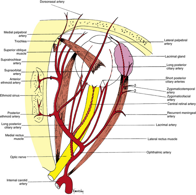
Anatomy of retrobulbar vessels ophthalmic artery (OA), temporal short... Download Scientific
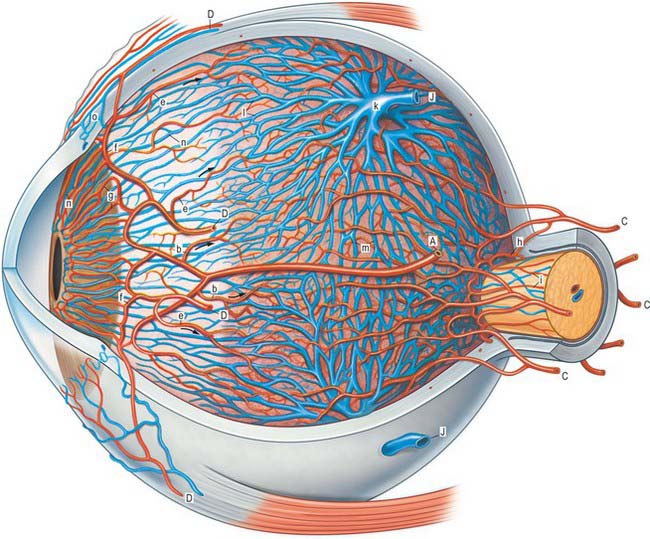
The short PCAs supply the following ( Figure 3 ): (1) the choroid as far as the equator, and (2) the overlying retina to a depth of about 130 μm, including the retinal pigment epithelium and up to the outer part of the inner nuclear layer.

The porcine short posterior ciliary arteries. Photograph showing the... Download Scientific
The ciliary muscle is an important part of the eye that contributes to a person's ability to view objects clearly at varying distances. [1] [2] [3] [4] Go to: Structure and Function The middle layer of the eyeball, called the vascular tunic, is composed of the choroid, ciliary body, and iris.

Schematic diagram The Arrangement of Nerve Fibers in the Retina and... Download Scientific
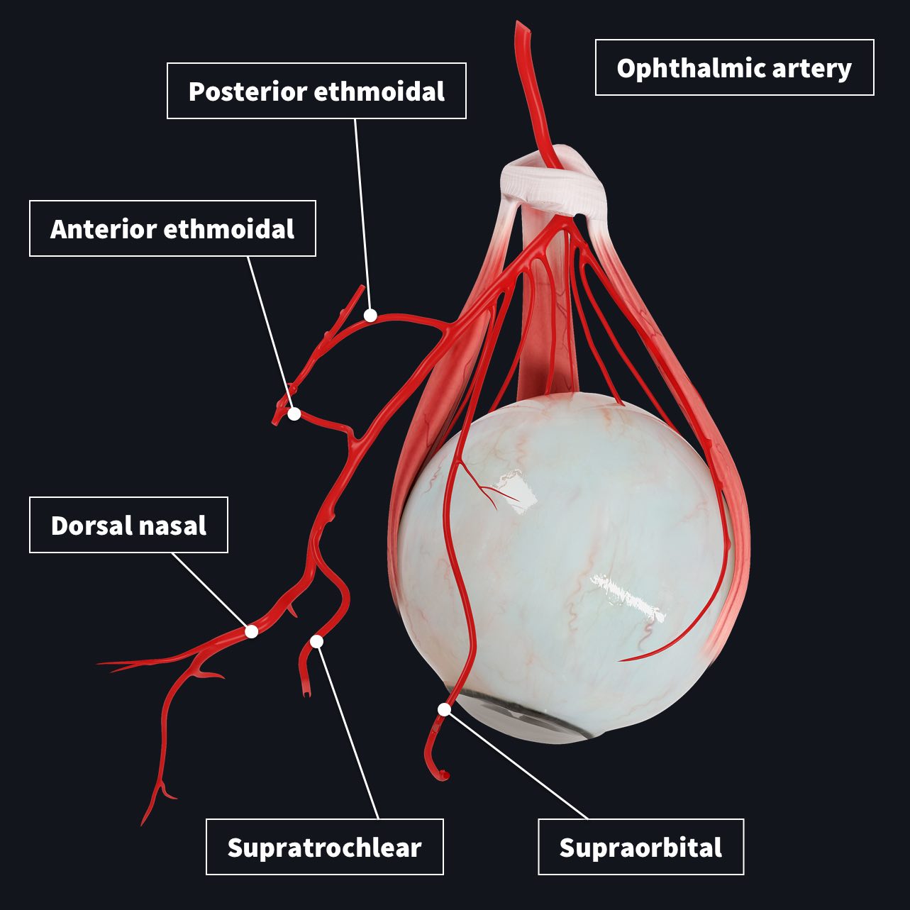
Short posterior ciliary arteries Long posterior ciliary arteries Muscular arteries Supraorbital artery Anterior ethmoidal artery Middle ethmoidal artery Posterior ethmoidal artery Medial palpebral arteries

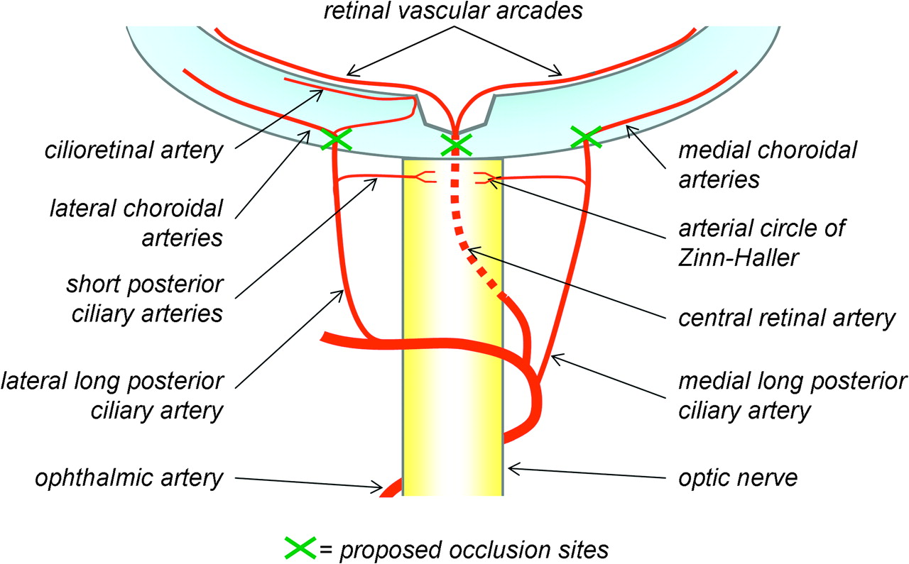
Schematic Drawing of the Ophthalmic Artery, Its Branches, and Possible... Download Scientific
The short posterior ciliary arteries arise from the ophthalmic artery. There are usually about seven arteries. Course The short posterior ciliary arteries extend anteriorly towards the eyeball and divide into 15-20 branches (Standring, 2016). They then pierce the sclera of the eyeball near the optic nerve. Branches
The eye Basicmedical Key
The posterior ciliary artery divides into the short and long posterior ciliary arteries that penetrate through the sclera and provide blood flow to the posterior uveal tract. Blood flow to the retina remains constant regardless of intraocular pressure, systemic blood pressure, and is independent of sympathetic autoregulation..

Temporal short posterior ciliary arteries (SPCA) identified by Esaote... Download Scientific
She described the development of the cilioretinal artery by the enlargement of an anastomosis of one of the posterior ciliary arteries with a small branch from the hyaloid artery on the disc. This anastomosis is located in man at the edge of the optic disc where traces of the short ciliary arteries enter the nerve near the lamina cribrosa in.

short posterior ciliary artery meddic
Purpose: Malfunction in peripapillary hemodynamics has been suggested to play a major part in the pathogenesis of primary open-angle glaucoma (POAG). The aim of this study was to determine whether topically applied brimonidine can influence blood hemodynamic characteristics associated with the perioptic short posterior ciliary arteries (SPCAs), central retinal artery (CRA), and choroidal.
Orbital Blood Supply Clinical Gate
The short posterior ciliary arteries are branches of the posterior ciliary arteries which are, in turn, branches of the ophthalmic artery. Each eye has multiple small short posterior ciliary arteries (16-20) which pierce the sclera adjacent to the optic nerve.
Vasculature of the eye Complete Anatomy
The ophthalmic artery has multiple branches which separate into two categories: orbital branches and optical branches. The orbital arteries include the ciliary arteries, central retinal artery, and muscular arteries. [1]
Orbital Blood Supply Basicmedical Key
Short posterior ciliary arteries, which penetrate the sclera around the optic nerve From: Slatter's Fundamentals of Veterinary Ophthalmology (Fourth Edition), 2008 Related terms: Axon Nerve Fiber Choroid Ischemic Optic Neuropathy Optic Canal Ciliary Arteries Long Posterior Ciliary Arteries Anterior Ciliary Arteries Optic Nerve Dog View all Topics
Arterial occlusion after scleral buckling British Journal of Ophthalmology
The posterior ciliary arteries are usually paired branches arising from the ophthalmic artery, one medial and one lateral, each giving off a number of branches that supply the uvea 1. Close to the optic nerve, are the short posterior ciliary arteries, usually numbering 16-20; these supply the posterior part of the choroid. Further from the.

Blood Vessels of the Eye Arizona RETINA Project
The meaning of SHORT POSTERIOR CILIARY ARTERY is any of 6 to 10 arteries that arise from the ophthalmic artery or its branches, pass to the posterior part of the eyeball while surrounding the optic nerve, and enter or divide into branches entering the sclera to supply the choroid and the ciliary processes.
