Figure 1 from Anatomy of the pleura reflection lines and recesses. Semantic Scholar

Anatomy of the pleura Osmosis
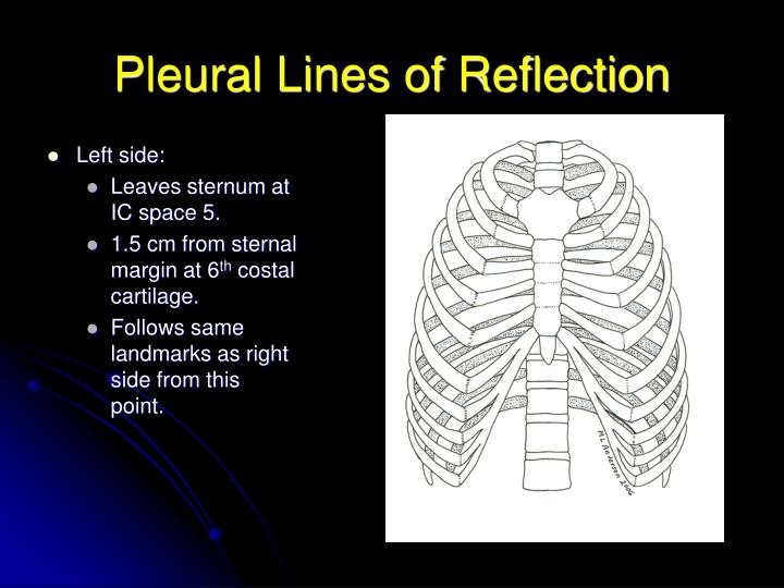
It is a sharp and thin border that overlays the pericardium that reaches the area where the parietal pleura reflects at the junction of the costal cartilages and the mediastinum (i.e. the costomediastinal line of pleural reflection). While this border is almost vertical on the right-hand side, on the left, it has a variable course.
PPT THORACIC CAVITY PowerPoint Presentation ID479328
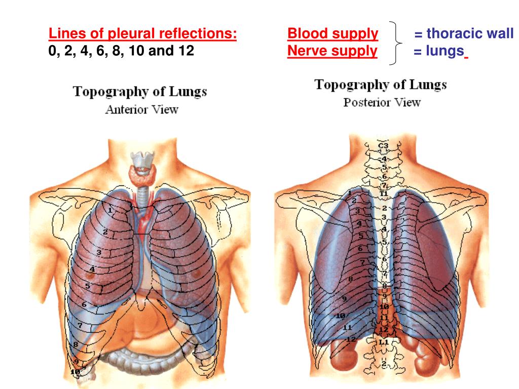
The lines of pleural reflection are formed by the parietal pleura as it changes direction (reflects) from one wall of the pleural cavity to another. The sternal lines of reflection are where the costal parietal pleura becomes continuous with the mediastinal pleura.
Lecture 3 lungs & pleura
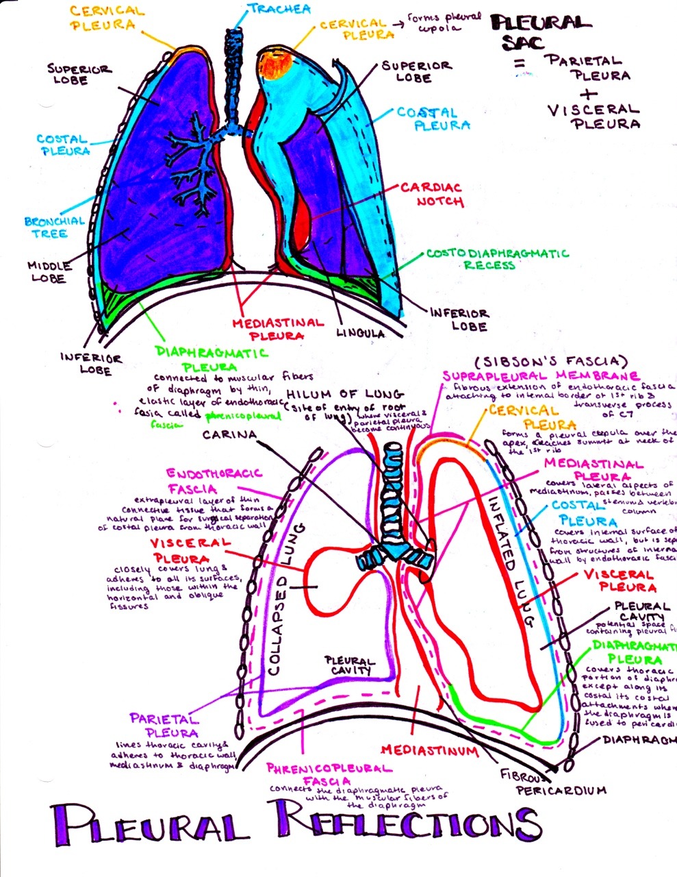
The pleura is a double-layered serous membrane that covers each lung and lines the thoracic cage. The outer layer ( parietal pleura) attaches to the chest wall. The inner layer (visceral pleura) covers the lungs, neurovascular structures of the mediastinum and the bronchi.

Visceral Pleura Definition Anatomy
The visceral and the mediastinal parietal pleurae are connected at the root of the lung ("hilum") through a smooth fold known as pleural reflections, and a bell sleeve -like extension of visceral pleura hanging under to the hilum is known as the pulmonary ligament .

Pleura Learn Surgery Online
Pleural space The pleural cavity surrounds the lungs in the thoracic cavity. There are two pleural cavities, one for each lung on the right and left sides of the mediastinum. Each pleural cavity and it's enclosed lung are lined by a serous membrane called pleura. The right and left pleural cavities are completely independent compartments.
5 The Pleura Radiology Key
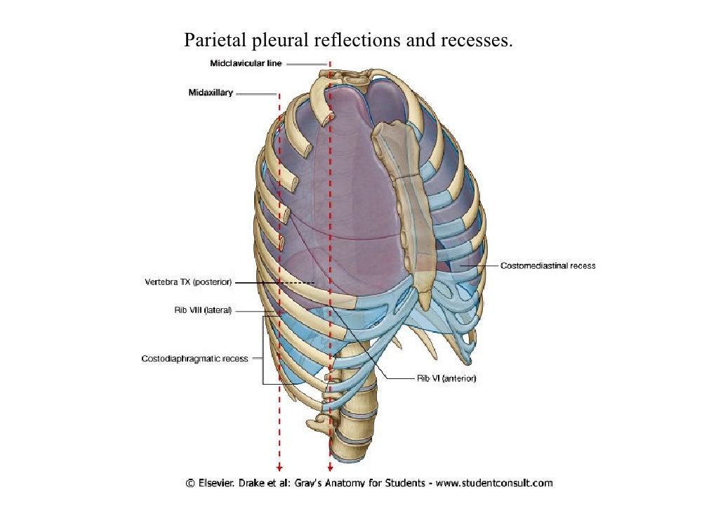
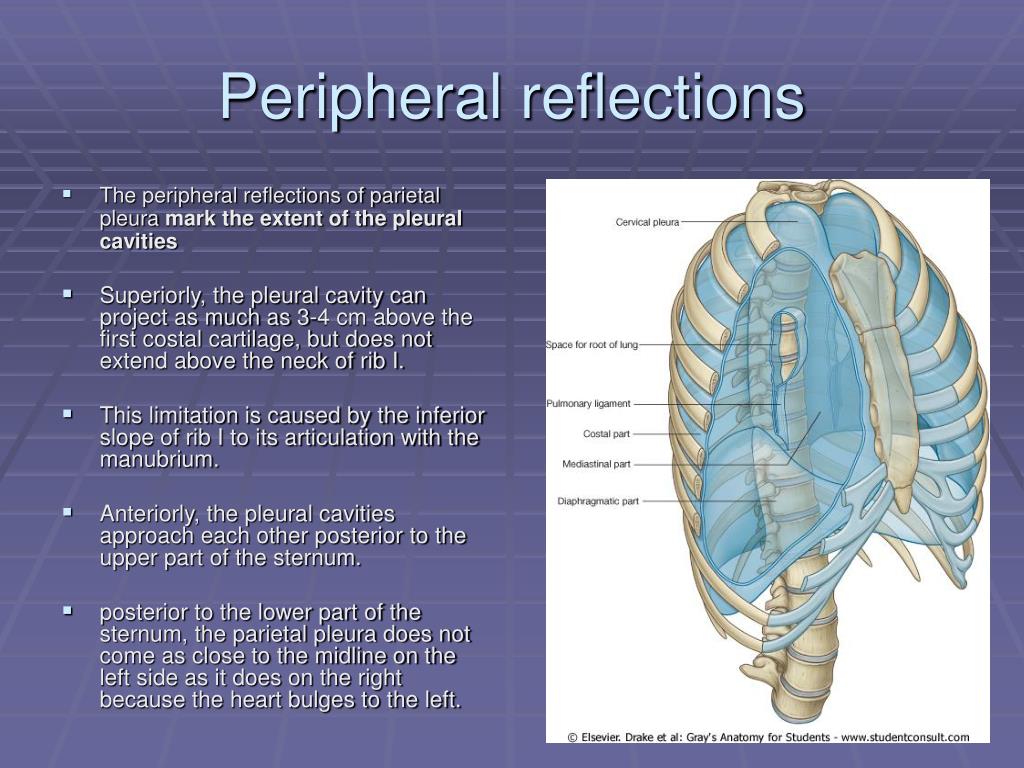
Reflections of the Pleura (pleural recesses, ligament and fascia):Commencing at the sternum, the pleura passes lateralward, lines the inner surfaces of the costal cartilages, ribs, and Intercostales, and at the back part of the thorax passes over the sympathetic trunk and its branches, and is reflected upon the sides of the bodies of the vertebr.
5 The Pleura Radiology Key
Function Related Conditions Frequently Asked Questions The pleura is a vital part of the respiratory tract. Its role is to cushion the lung and reduce any friction that may develop between the lung, rib cage, and chest cavity. Each pleura (there are two) consists of a two-layered membrane that covers each lung.
PPT Sternal angle PowerPoint Presentation, free download ID957362
A pleura is a serous membrane that folds back on itself to form a two-layered membranous pleural sac. The outer layer is called the parietal pleura and attaches to the chest wall. The inner layer is called the visceral pleura and covers the lungs, blood vessels, nerves, and bronchi.

Anatomy of the pleura Osmosis
Figure 1. A Layers of the thoracic wall. B Anterolateral view of the pleurae with the ribs partially removed and the parietal pleura removed on the right lung. C Anterolateral view of the surfaces of the parietal pleura, with the ribs partially removed and the left lung removed completely. Figure 2.
PPT Trachea and lungs PowerPoint Presentation, free download ID4779845
Pleural Cavity The pleural cavity is a potential space between the parietal and visceral pleura. It contains a small volume of serous fluid, which has two major functions. It lubricates the surfaces of the pleurae, allowing them to slide over each other.
hanson's anatomy — Pleural reflections
The lines of pleural reflection are formed by the parietal pleura as it changes direction (reflects) from one wall of the pleural cavity to another. The sternal lines of reflection are where the costal parietal pleura becomes continuous with the mediastinal pleura.

Pleura Anatomy 2 (Pleural reflections & Pleural Recess) YouTube
The lines of pleural reflection outline where parietal pleura abruptly changes direction as it passes from one wall of the pleural cavity to another. Right and left parietal pleura reflect in an asymmetric manner due to the presence of the heart.

PPT THORACIC CAVITY PowerPoint Presentation, free download ID479328
At the inferior aspect of the root, the reflection of the mediastinal pleura over the visceral pleura continues as a bilayered sheet of both visceral and parietal pleura (total of four layers) in the front and back of the hilar plane to form the inferior pulmonary ligament (also known as the triangular ligament), that is carried downward and mor.

Anatomy of the Pleura Thoracic Surgery Clinics
Knowledge of the anatomy of the lines of pleural reflection, triangular ligaments, and pleural recesses is important to thoracic surgeons because their anatomic areas are used daily for radiographic interpretation as well as for the performance of procedures such as chest tube insertion, thoracentesis, and pericardiocentesis. Their knowledge is.
Thorax Basicmedical Key
Abstract. Knowledge of the anatomy of the lines of pleural reflection, triangular ligaments, and pleural recesses is important to thoracic surgeons because their anatomic areas are used daily for.

Figure 1 from Anatomy of the pleura reflection lines and recesses. Semantic Scholar
Pleura: Anatomy. The pleura is a serous membrane that lines the walls of the thoracic cavity and the surface of the lungs. This structure of mesodermal origin covers both lungs, the mediastinum, the thoracic surface of the diaphragm, and the inner part of the thoracic cage. The pleura is divided into a visceral pleura and parietal pleura.
