Lesson 3 Onion Dissection & “Look at the Plant Cells” Rs' Science
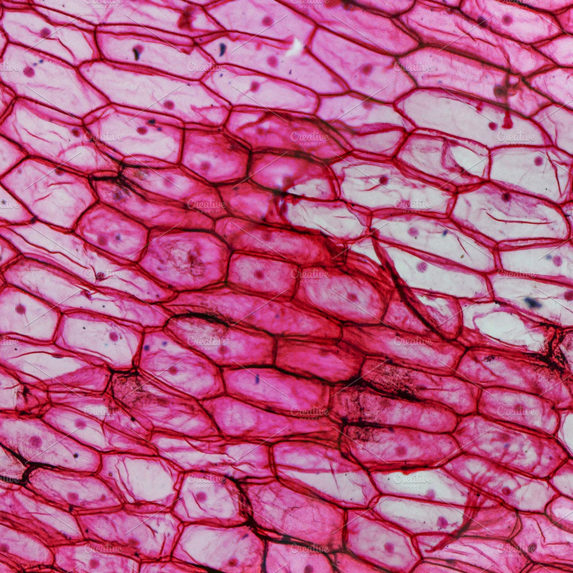
Onion cells HighQuality Nature Stock Photos Creative Market
The graphic shows that the majority of the cells are in interphase, and students are asked to calculate the percentage. The cells are drawings, but they are meant to model real cells in mitosis. The graphic shown below compares drawings to real cells. Part of my biology class is helping students navigate diagrams and data.

Onion peel cell diagram Cell diagram, Museum exhibition design, Exhibition design
Figure 10.1.5 10.1. 5: A micrograph of a cell nucleus. The nucleolus (A) is a condensed region within the nucleus (B) where ribosomes are synthesized. The nucleus is surrounded by the nuclear envelope (C). Just oustide the nucleus, the rough endoplasmic reticulum (D) is composed of many layers of folded membrane.

onion cells Google Images Biology art, Microscopy art, Nature inspiration
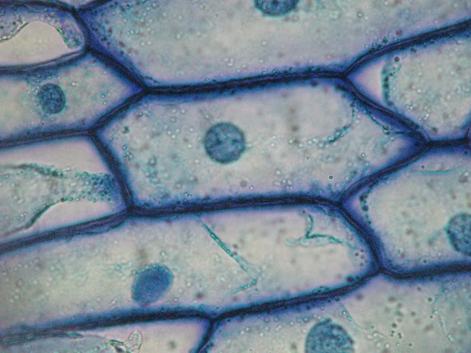
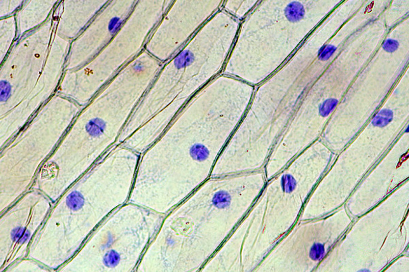
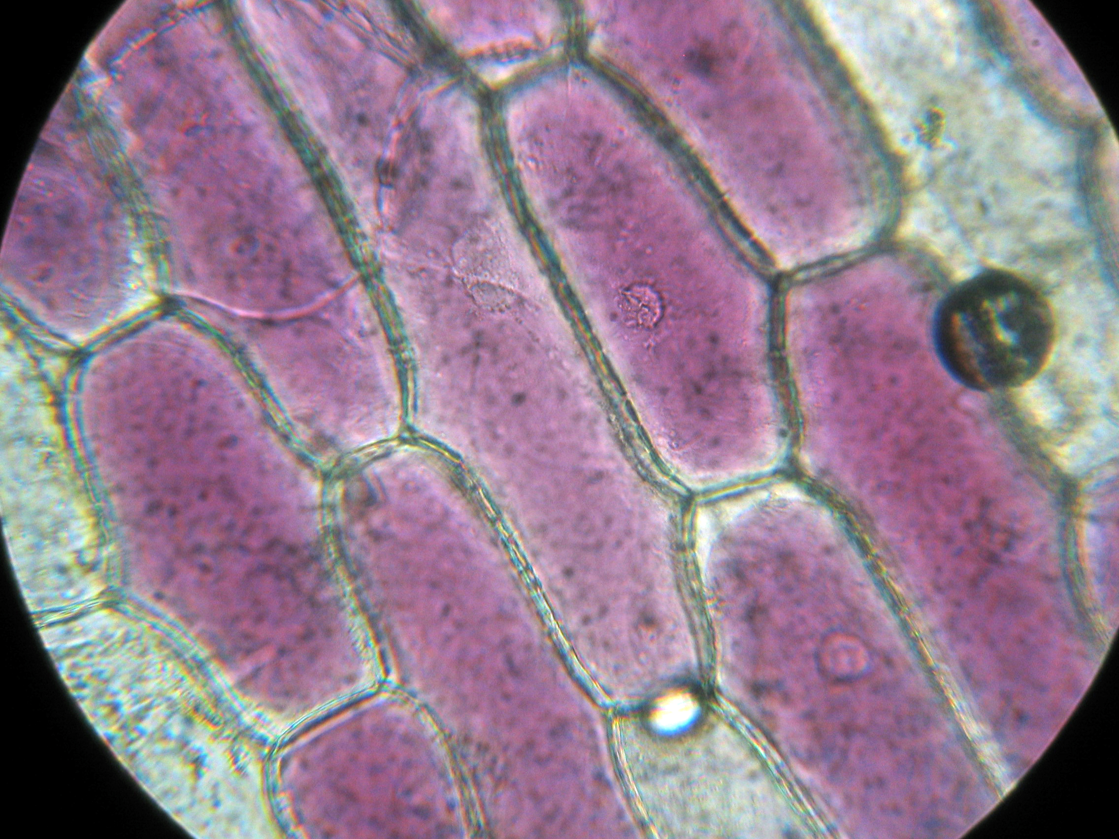
Figure 10.3.1.1 10.3.1. 1: Cells in an onion root in interphase and prophase. Cell A has a large, dark nucleolus surrounded by greyish material (chromatin) that is enclosed within the nuclear membrane. A cell wall makes a box around each cell and the plasma membrane would be located just inside this box, though we cannot easily see it.
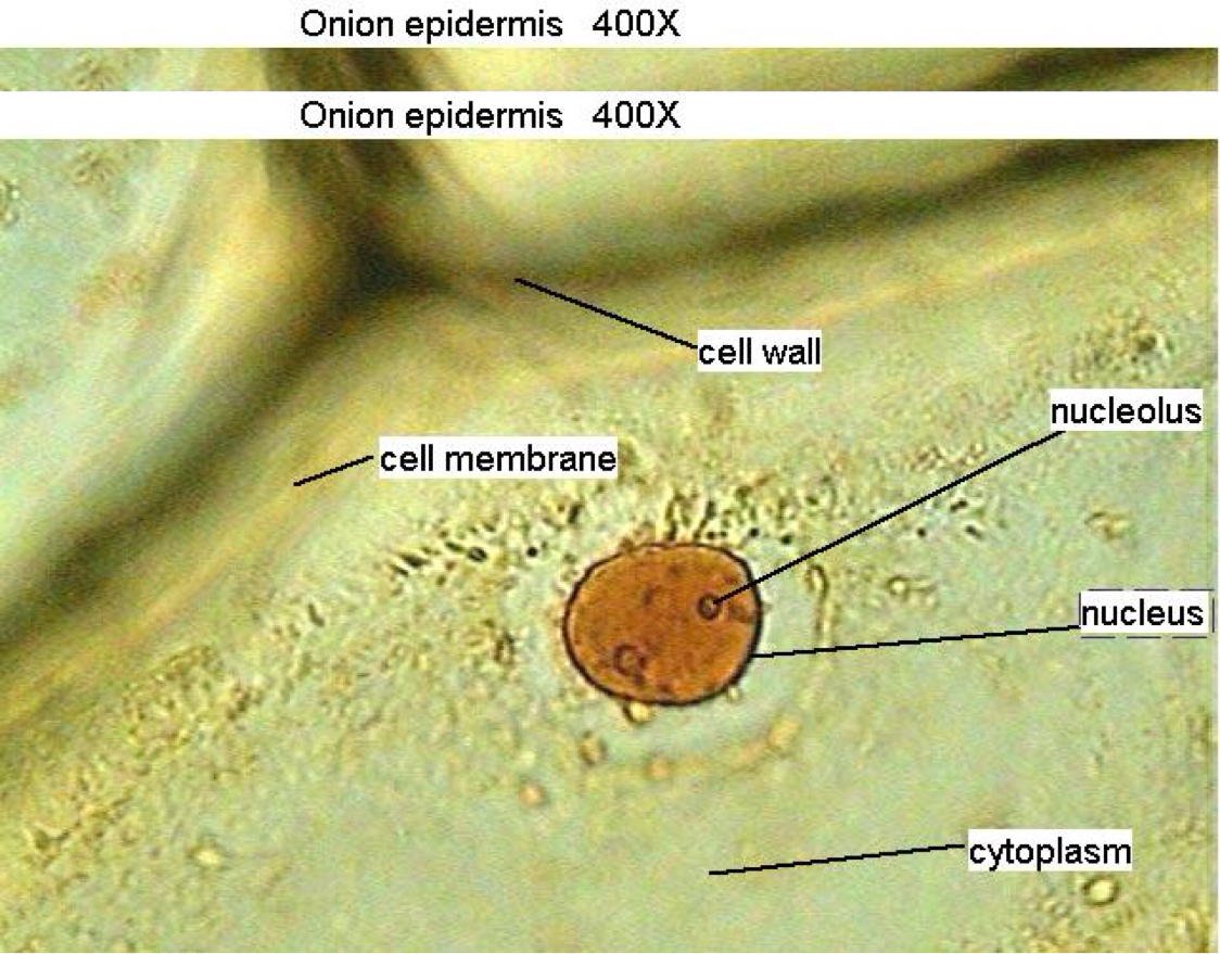
Onion Cell Under Microscope Labeled Drawing apostolicavideo
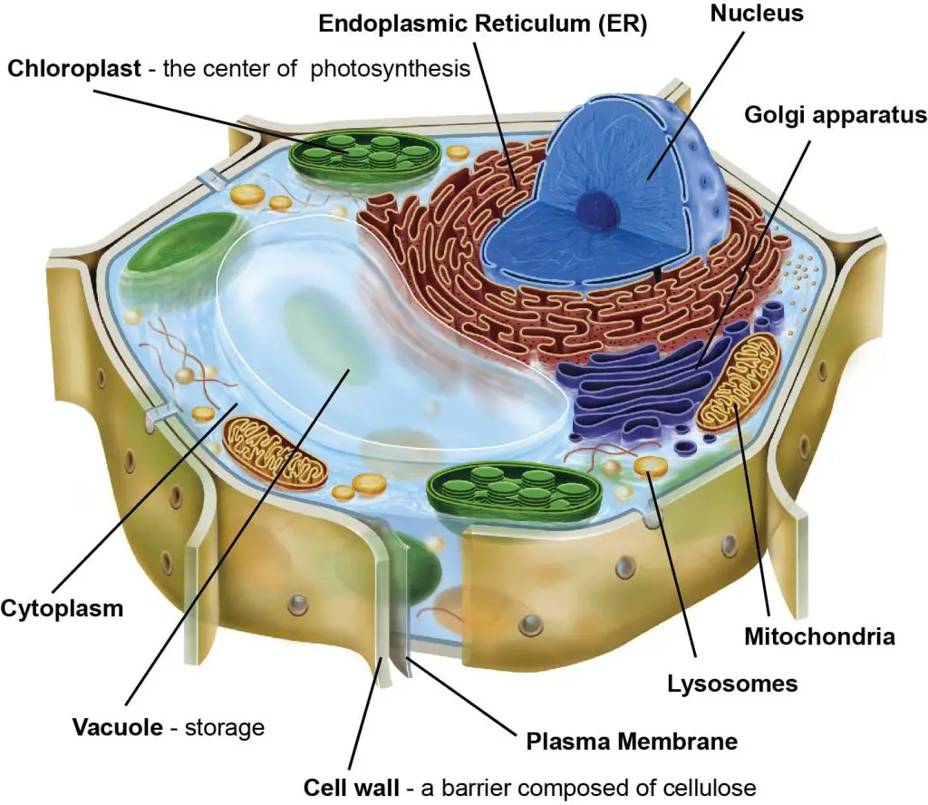
To answer your question, onion cells (you usually use epithelial cells for this experiment) are 'normal' cells with all of the 'normal' organelles: nucleus, cytoplasm, cell wall and membrane, mitochondria, ribosomes, rough and smooth endoplasmic reticulum, centrioles, Golgi body and vacuoles.
Onion cells slide (80× magnification) microbiology
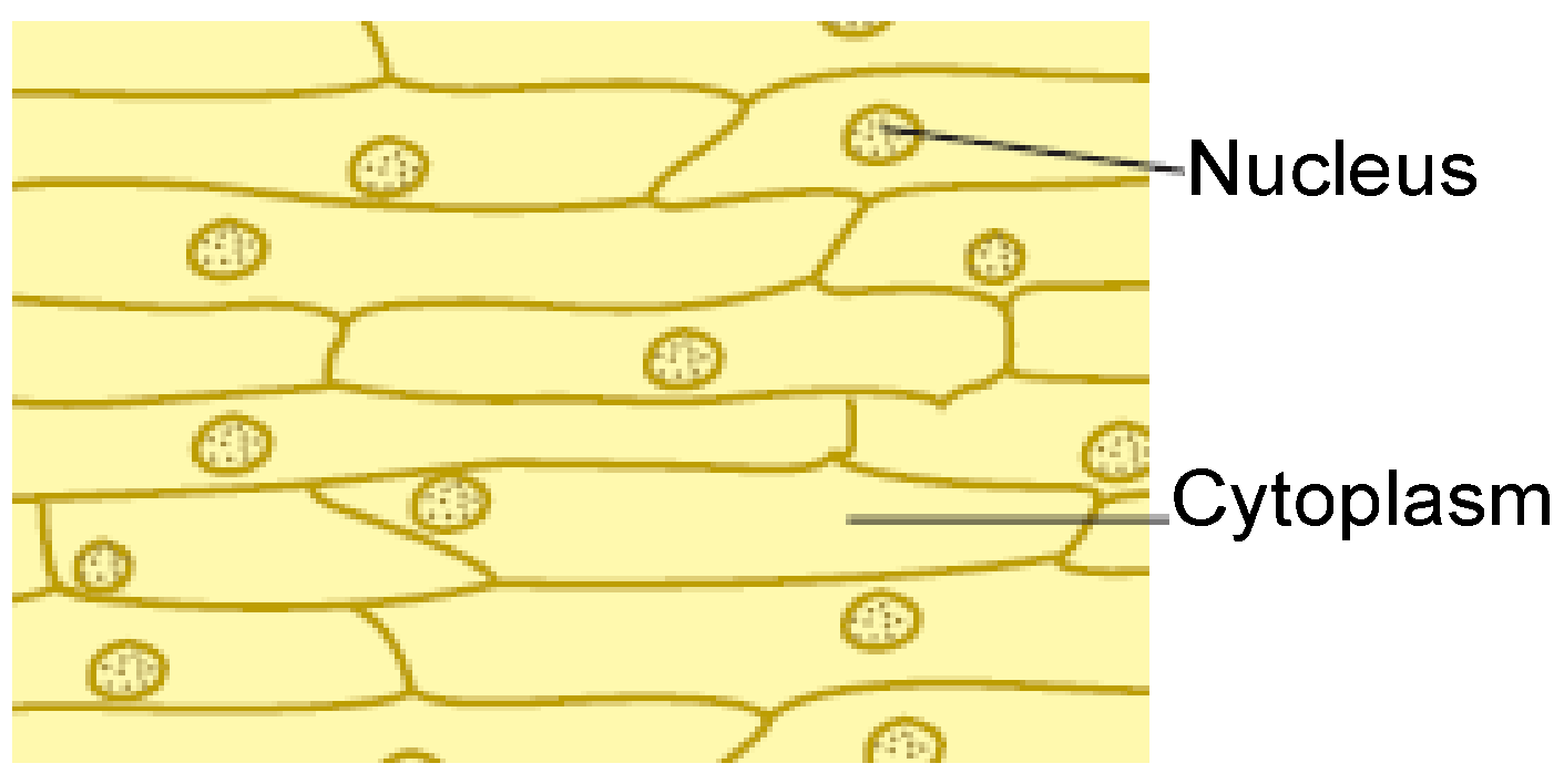
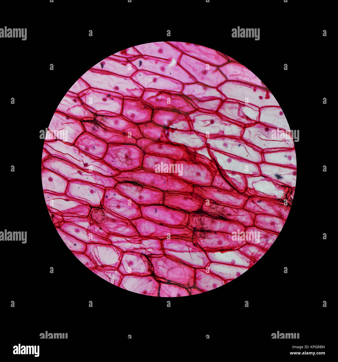
The Onion and Cheek Cell Lab Background: Onion tissue provides excellent cells to study under the microscope. The main cell structures are easy to see when viewed with the microscope at low power. For example, you will observe a large circular nucleus in each cell, which contains the genetic material for the cell.
Beautiful World Onion cells
The cells are the functional units of all organisms. All cells arise from preexisting cells. All cells share common features such as having a plasma membrane, a cytoplasm, DNA, and ribosomes. A plasma membrane is a phospholipid bilayer that surrounds the cell. This thin and fluid layer around the cells serves to isolate the cell's contents.
Onion Cell
The cell wall tends to give plant cells a boxy, rigid structure. Figure 3.8.1 3.8. 1: Elodea leaf cells. The most obvious of the membrane-bound organelles you will see are the chloroplasts. These numerous, green, disc-like structures are responsible for doing photosynthesis, making food for the plant.

draw the figure of an onion peel showing cell Brainly.in
The Onion Peel Cell Experiment is a popular and educational activity used to observe and understand the structure of plant cells. This experiment focuses on the onion, a eukaryotic plant known for its multicellular composition. As we delve into this experiment, we explore the essential components that make up a cell, the building blocks of life.

Biology LectureHub
What do onion cells look like under the microscope? Studying cell tissues from an onion peel is a great exercise in using light microscopes and learning about plant cells, since onion cells are highly visible under a microscope, especially when stained correctly.

Nuclei of onion cells Cell, Nucleus, Homeschool
An onion is a multicellular (consisting of many cells) plant organism. As in all plant cells, the cell of an onion peel consists of a cell wall, cell membrane, cytoplasm, nucleus and a large vacuole. The nucleus is present at the periphery of the cytoplasm. The vacuole is prominent and present at the centre of the cell.
Mitochondria In Onion Peel Cell Diagram Debsartliff
Next, prepare a slide of plant cells by placing five drops of distilled water into a clean watch glass. Then, with forceps, take a thin strip of onion and place it into the water. Apply 5 drops of safranin solution to another watch glass. Now using a pair of forceps transfer the piece of onion from the distilled water into the safranin.
FileRed Onion Cells.JPG Wikipedia
Fill out the Venn diagram below to show the differences and similarities between the onion cells and the Elodea cells. This page titled 1.5: Cells is shared under a CC BY-SA 4.0 license and was authored, remixed, and/or curated by Susan Burran and David DesRochers.

Epidermal onion cells under a microscope. Plant cells appear polygonal from the Cell diagram
Objective The main objective of performing the onion peel cell experiment is to observe the arrangement and structural components of the onion epidermis. The following facts about the onion peel cell experiment play a significant role in educating students: The epidermis of the onion bulb is a single layer of tissue that is easy to separate.
Onion cells microscope hires stock photography and images Alamy
Epidermal cells of the onion bulb assemble a pectin-rich polylamellate outer wall that is readily isolated as centimeter-scale strips by peeling, lending itself to wide-ranging techniques for structural analysis, including spectroscopy, mechanics, and microscopy. Extending this toolkit, Nicolas et al., in a new study in this issue of Current.

onion cell diagram Laceness
Onion Cells Under a Microscope ** Requirements, Preparation and Observation The bulb of an onion is formed from modified leaves. While photosynthesis takes place in the leaves of an onion containing chloroplast, the little glucose that is produced from this process is converted in to starch (starch granules) and stored in the bulb.
Lesson 3 Onion Dissection & “Look at the Plant Cells” Rs' Science
Updated July 11, 2019 By Peg Robinson Onions have a long history of human use, originating in southwestern Asia but having since been cultivated across the world. Their strong odor — actually a defense mechanism — and unique structure belie a complex internal makeup, composed of cell walls, cytoplasm, and the vacuole.
