Brain Atlas of human anatomy with MRI eAnatomy

MRI anatomy brain axial image 18 Brain anatomy, Mri brain, Brain scan
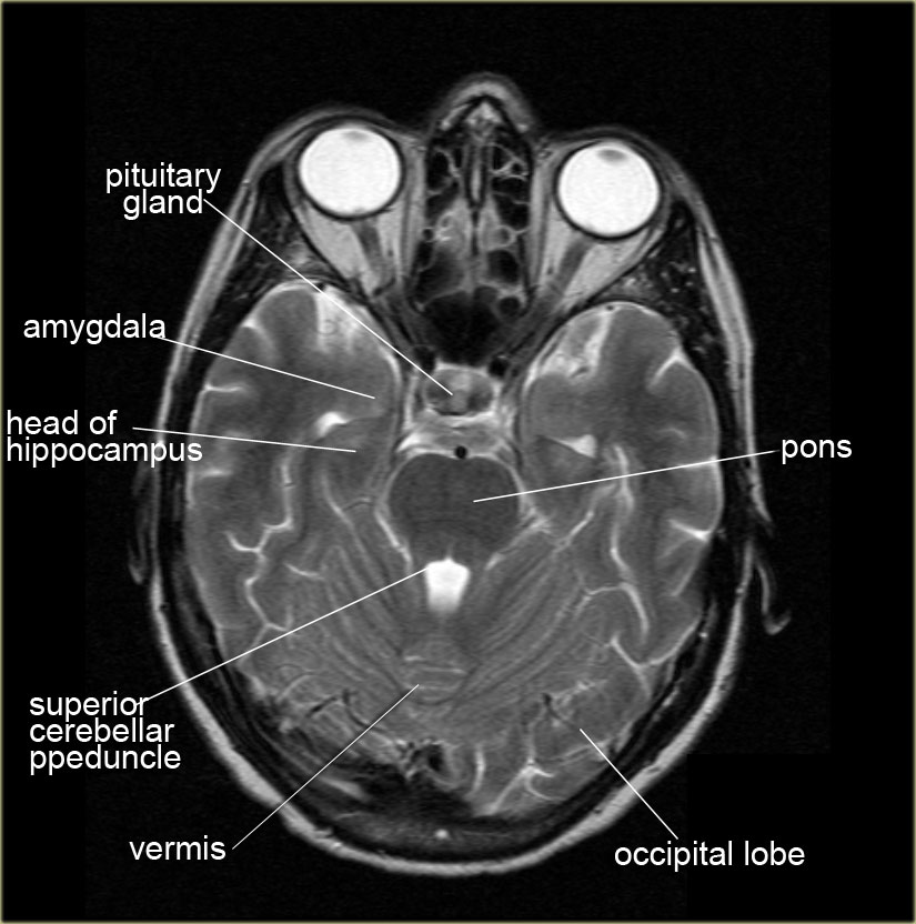
Anatomy of the Brain The advent of high-resolution computed tomography (CT) and magnetic resonance imaging (MRI) scanners has allowed the fine anatomic structure to be seen in detail.

Brain MRI
A brain (head) MRI scan is a painless test that produces very clear images of the structures inside of your head — mainly, your brain. Healthcare providers use brain MRIs to evaluate, diagnose and monitor several different medical conditions that affect your brain or other structures in your head.

The Radiology Assistant Brain Anatomy
It is the main method to investigate conditions such as multiple sclerosis and headaches, and used to characterize strokes and space-occupying lesions. Reference article This is a summary article; we do not have a more in-depth reference article. Summary indications confirmation of stroke assessment of intracranial tumor chronic headache

Approach to MRI brain
Brain magnetic resonance imaging (MRI) is a common medical imaging method that allows clinicians to examine the brain's anatomy(1). It uses a magnetic field and radio waves to produce detailed images of the brain and the brainstem to detect various conditions(2).
Functional Brain Anatomy Radiology Key
Anatomy of the brain (MRI) - cross-sectional atlas of human anatomy Antoine MICHEAU, MD , Denis HOA, MD Authors affiliations Publication date: Aug 25, 2008 | Last update: Oct 5, 2022 https://doi.org/10.37019/e-anatomy/163 ISSN 2534-5079

Mri Brain Anatomy Radiology Masterclass ANATOMY STRUCTURE
Edit article Citation, DOI, disclosures and article data This article lists a series of labeled imaging anatomy cases by body region and modality. Brain CT head: non-contrast axial CT head: non-contrast coronal CT head: non-contrast sagittal CT head: non-contrast axial with clinical questions CT head: angiogram axial CT head: angiogram coronal
Functional Brain Anatomy Radiology Key
A head MRI is a useful tool for detecting a number of brain conditions, including: aneurysms, or bulging in the blood vessels of the brain multiple sclerosis spinal cord injuries.
Brain Anatomy On Mri Anatomical Charts & Posters
MSK MRI ATLAS illustration by Kate Stevens Atlas: Hip Atlas: Thigh Lecture: Basic hip: Atlas: Knee Atlas: Lower leg Lecture: Meniscus Lecture: Patellar Measurements Cases: Basics J. Harter Cases: Lateral corner S. Honowitz Cases: Medial corner S. Honowitz: Atlas: Ankle Lecture: Chopart (RSNA award)

resonance image (MRI) of the brain ODC
Deep spaces of the head and neck - annotated MRI Case contributed by Jeffrey Hocking Diagnosis not applicable Share Add to Citation, DOI, disclosures and case data Diagram Axial MRI sequence illustrating the deep spaces of the head and neck. 10 articles feature images from this case 165 public playlists include this case

Normal Brain MRI Anatomy Neuroradiology Made simple YouTube
This video shows the appearance of the anatomical structures of the brain on a Magnetic Resonance Imaging.It aims to complement your understanding of neuroan.
MRI Brain Anatomy
Due to the large number of muscle structures of the head and neck, we had to use several groups of muscles: muscles of the face, tongue, pharynx, larynx, neck, back and masticator muscles. Anatomy of the oral cavity, nose and sinus of the face : coronal slice with anatomical structures labeled

Brain Atlas of human anatomy with MRI eAnatomy
MRI is used to analyze the anatomy of the brain and to identify some pathological conditions such as cerebrovascular incidents, demyelinating and neurodegenerative diseases. Moreover, the MRI can be used for examining the activity of the brain under specific activities (functional MRI - fMRI).

MRI anatomy brain axial image 6 Mri brain, Brain anatomy, Mri
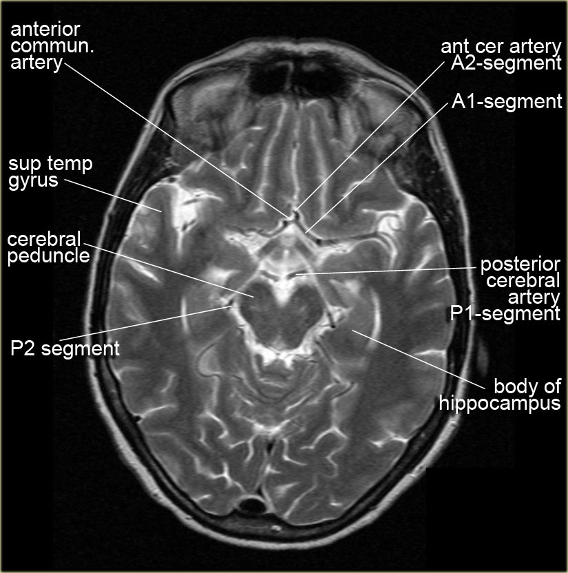
Also called ambient cistern is a cistern of the subarachnoid space between the posterior end of the corpus callosum and the superior surface of the cerebellum. It is sometimes defined as including the quadrigerminal cistern. On the left a coronal view of the segments of the middle cerebral artery. Horizontal M1-segment

Brain Anatomy On Mri Anatomical Charts & Posters
Normal Brain: Normal Anatomy in 3-D with MRI/PET (Javascript) Atlas of normal structure and blood flow Top 100 Brain Structures Can you name these brain structures? Normal aging: structure and function Normal aging: structure and function Normal aging: coronal plane Vascular anatomy Cerebrovascular Disease (stroke or "brain attack"):

Mri Anatomy Of Brain ANATOMY
Reading time: 31 minutes Normal chest x ray Radiological anatomy is where your human anatomy knowledge meets clinical practice. It gathers several non-invasive methods for visualizing the inner body structures. The most frequently used imaging modalities are radiography ( X-ray ), computed tomography ( CT) and magnetic resonance imaging ( MRI ).
The Radiology Assistant Brain Anatomy
The MRI is a particularly powerful exam for studying structures such as diencephalon, mesencephalon (mid brain), pons, myelencephalon (medulla oblongata, bulb) and spinal cord. The vertical left menu provides reference images on coronal and sagittal views of the brain, with anatomical schemas based on a three dimensional (3D) model.
