Transmission electron micrograph of an animal cell Stock Image G450/0052 Science Photo Library
Transmission electron micrograph of animal cell Stock Image G450/0051 Science Photo Library
Electron micrographs of eukaryotic animal cells. Figure 4.24 TEM of a pancreas acinar (exocrine) cell. Figure 4.25 TEM of part of a eukaryotic cell. Figure 4.26 TEM showing a large and active Golgi apparatus. Figure 4.27 This electron micrograph shows the close relationship between rER and the Golgi apparatus. Figure 4.28 TEM of a mitochondrion.

Solved label the ectron micrograph of an animal cell.
EM tomography. Spherical protein shells of the hepatitis B virus are preserved in a thin film of ice (A) and imaged in the transmission electron microscope. Thousands of individual particles were combined by EM tomography to produce the three-dimensional
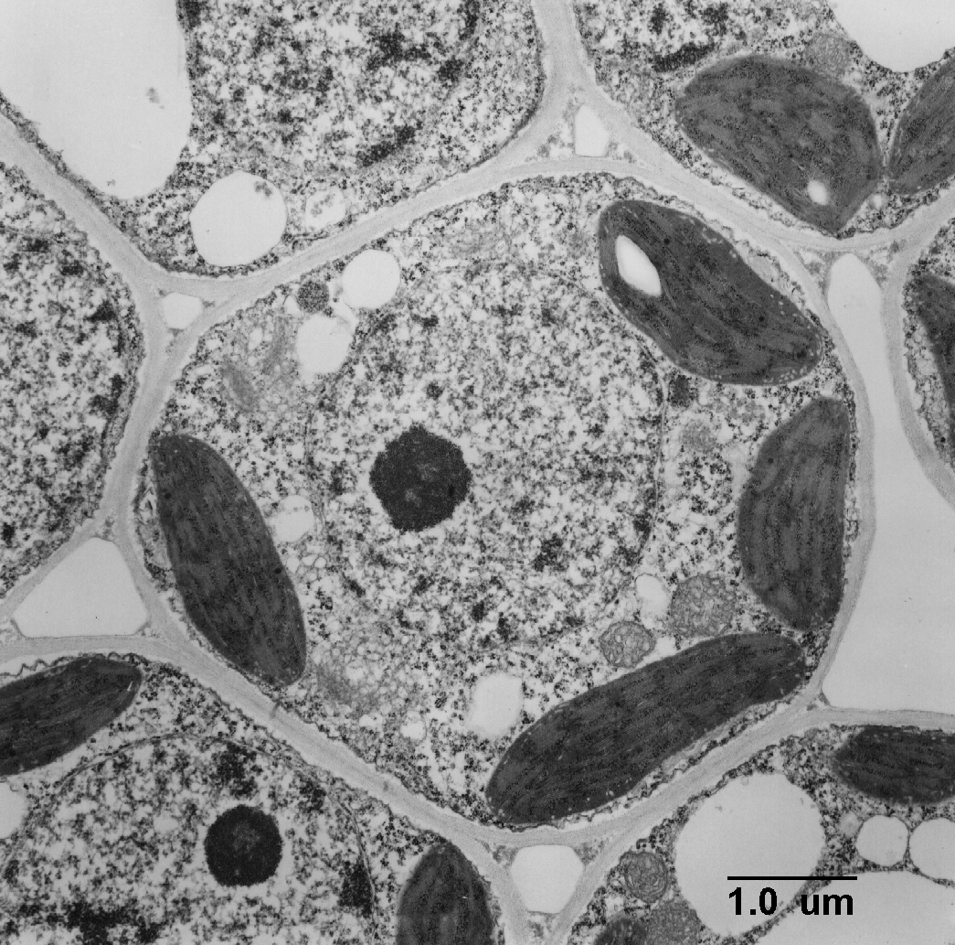
TEM of Plant Cell
Electron Microscopy of Animal Cells Organelles under the electron microscope There are two types of electron microscope Transmission electron microscopes (TEMs) Scanning electron microscopes (SEMs) Transmission Electron Microscopes TEMs use electromagnets to focus a beam of electrons This beam of electrons is transmitted through a thin specimen
Unit 7 Microscopes & Cells Grade 6 Science
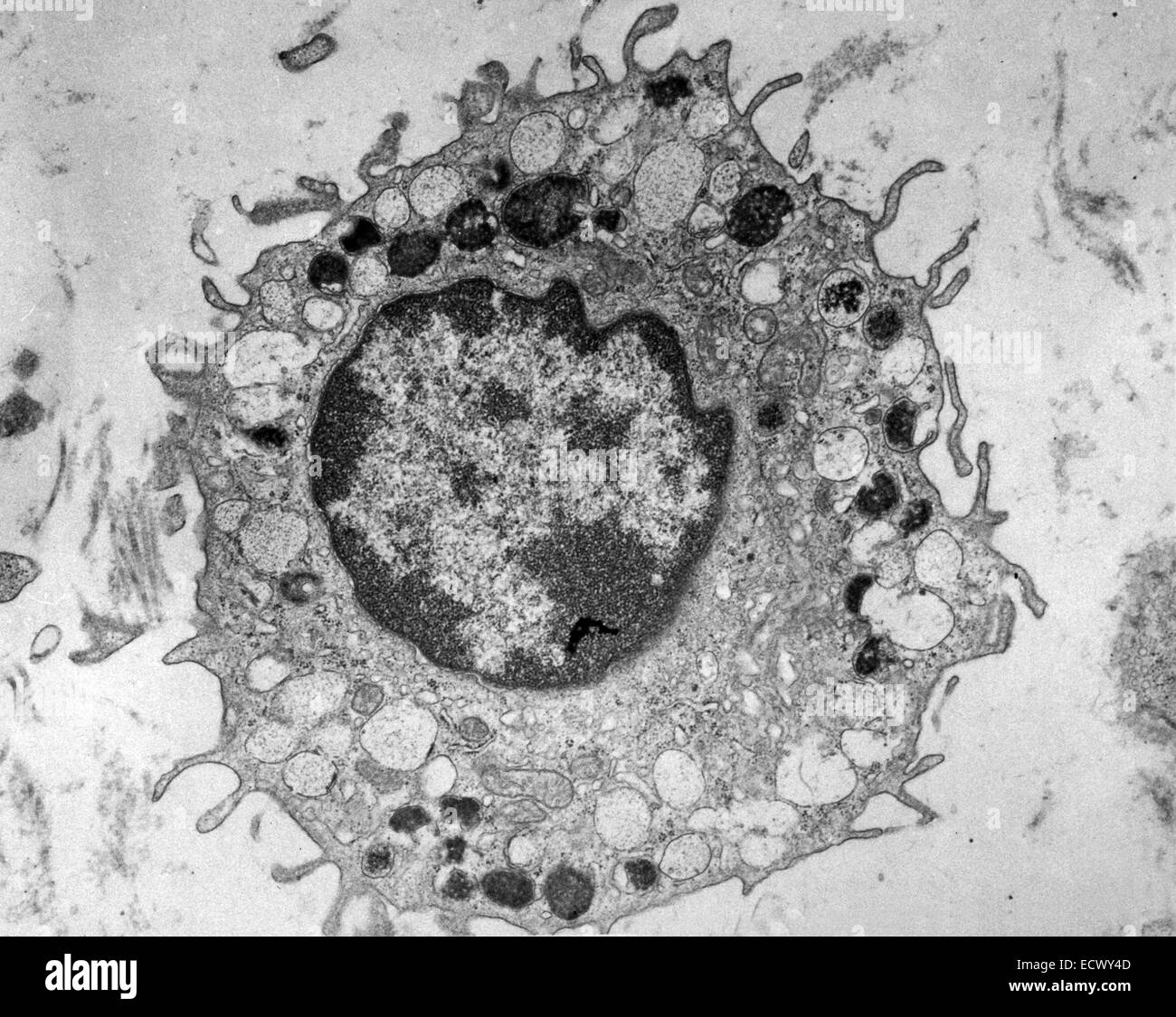
Figure 2 (a) A transmission electron microscope. (b) A transmission electron micrograph of a frog leukocyte (white blood cell). The nucleus and nucleolus (Section 4.3), mitochondria (Section 4.10) and Golgi apparatus (Section 4.7) can be seen. The dark area of the nucleus contains densely packed DNA. Show description.
Images 01. Introduction and Terminology Basic Human Anatomy
In an electron micrograph of leaf cells from a quiescent desiccated dessert plant, Selaginella lepidophylla,. In animal cells they participate in spindle fiber formation during mitosis and are the point from which microtubules radiate thorough the cell to help form and maintain its shape. These structures do not involve axonemes.
.jpg)
Year 11 Bio. Key Points February 2013
A microscope is an instrument that magnifies objects otherwise too small to be seen, producing an image in which the object appears larger. Most photographs of cells are taken using a microscope, and these pictures can also be called micrographs. From the definition above, it might sound like a microscope is just a kind of magnifying glass.

clothes and stuff online Animal Cell Electron Micrograph
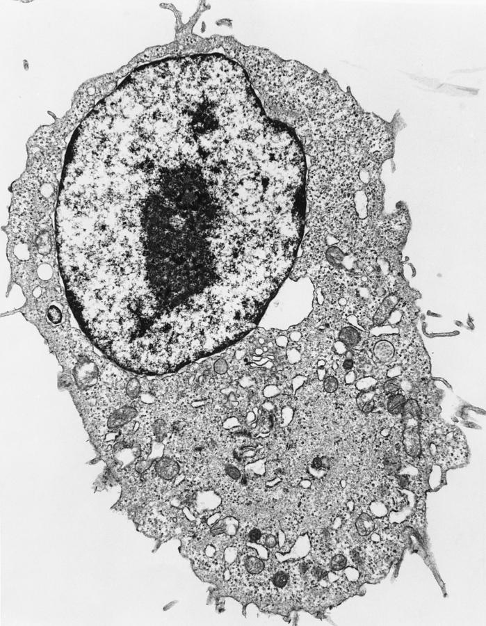
Animal cell. Transmission electron micrograph of a mammalian tissue culture cell. Taking up most of the cell is the nucleus, where genes are stored in the form of chromosomes. The dark zone (centre right) in the nucleus is the nucleolus. This is the most active part of the nucleus, and contains unravelled chromosomes involved in making protein.
Animal Cell Electron Microscope Fun Images
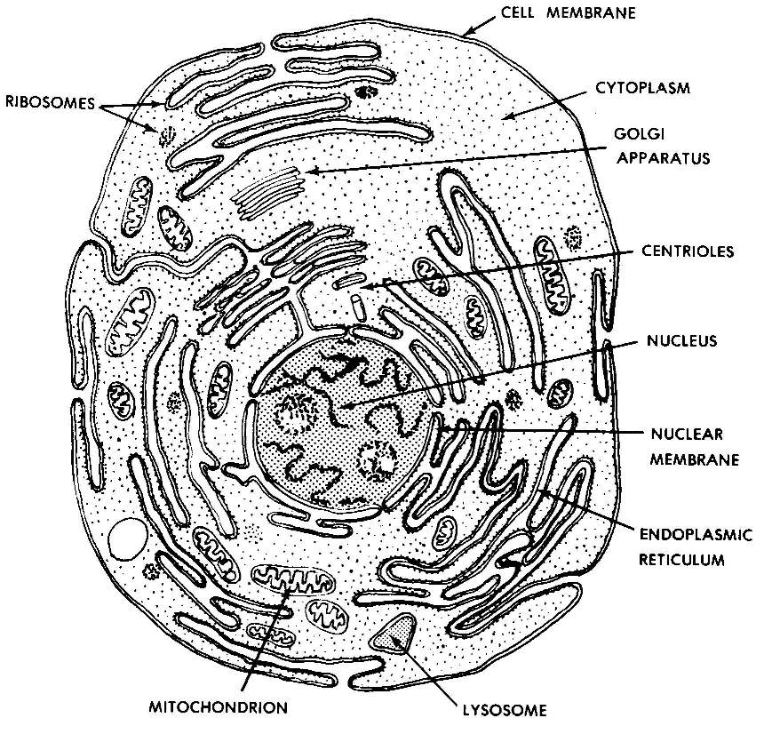
Animal Cell Structure Animal cells are typical of the eukaryotic cell, enclosed by a plasma membrane and containing a membrane-bound nucleus and organelles. Unlike the eukaryotic cells of plants and fungi, animal cells do not have a cell wall.
Electron micrograph of mammalian cell Stock Photo Alamy
The only structures found in animal cells but not plant cells are the centrioles and microvilli. Plant cells also have additional structures: the cellulose cell wall, large permanent vacuoles and chloroplasts. The ultrastructure of an animal cell shows a densely packed cell - the ER and RER and ribosomes form extensive networks throughout the.
Electron Micrograph Of Eukaryotic Animal Cell / What Is A Diagram Of A Plant And Animal Cell
Figure 3 Electron micrograph showing two cells of the bacterial species Neisseria meningitidis immunolabelled with a primary antibody that recognises an oligosaccharide, followed by a secondary antibody that has been coupled to individual 15 nm gold particles. Note that the cytoplasm has uneven electron density.. In animal cells.

clothes and stuff online Animal Cell Electron Micrograph
In this video lecture, students will learn how a typical animal cell looks like an electron microscope. The students will also learn how to label different s.

Cell Theory Introducing the Cell
It is responsible for controlling the cell. 2.3.3 Identify structures from 2.3.1 in electron micrographs of liver cells. Figure 2.3.2 - Electron micrograph of an animal cell 2.3.4 Compare prokaryotic and eukaryotic cells. Prokaryotic cells have naked DNA which is found in the cytoplasm in a region named the nucleoid.
Transmission electron micrograph of an animal cell Stock Image G450/0052 Science Photo Library
3 The Plasma Membrane 3.1 How substances move across the Plasma Membrane 4 The Cytoplasm 4.1 a) Cytosol 4.2 b) Cell inclusions 4.3 c) Organelles 4.3.1 Ribosomes 4.3.2 Endoplasmic reticulum 4.3.3 Mitochondria 4.3.4 Golgi Apparatus 4.3.5 Lysosomes 4.4 d) Microfilaments And Microtubules 5 The Nucleus 5.1 Chromosomes 6 Cell Division
Animal Cell Tem Labeled / consider the animal cell. Which organelle is labeled I and Give
Animal cell. Transmission electron micrograph of part of a mouse liver cell. In the lower frame is the cell nucleus, bound by a delicate nuclear membrane. Holes in the membrane (at right, for example) allow large molecules to pass out into the cell cytoplasm. The dark grey circles above the nucleus are mitochondria, where fats and sugars are.

Hepatocyte (liver cell) various organelles including the nucleus, lysosomes, mitochondria and
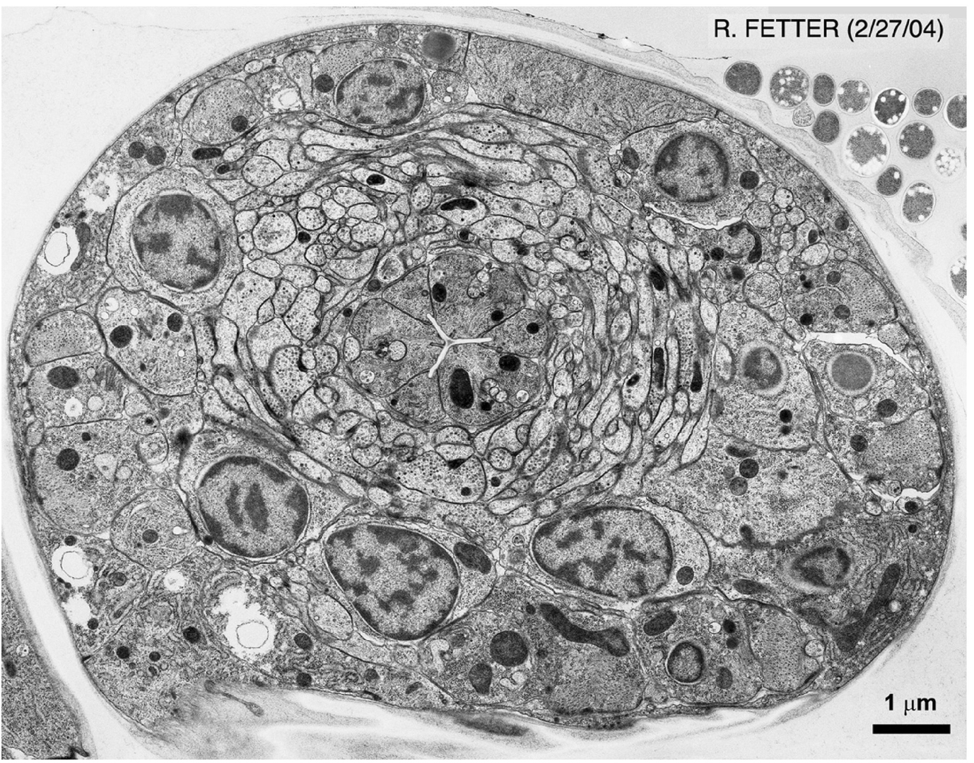
Prokaryotic cells like bacteria have no nucleus and fewer organelles than eukaryotic cells, which form plants and animals. Electron Micrographs Basic Understanding. An electron micrograph is a type of image created using electron microscopy. It provides high-resolution images allowing for detailed study of cell structures, including cell.

Electron Micrograph Of A Plant Cell
Electron microscopy (EM) uniquely visualizes cellular structures with nanometre resolution. However, traditional methods, such as thin-section EM or EM tomography, have limitations in that they.
