Parts of a Compound Microscope Labeled (with diagrams) Medical Pictures and Images (2023

Microscope Diagram Tim's Printables
Fluorescence microscopes: These use fluorescent dyes to highlight specific structures or molecules in a sample and are commonly used in biological research. X-ray microscopes: These use X-rays to produce images of the internal structure of samples and are often used to study materials and biological specimens.
Monday September 25 Parts of a Compound Light Microscope
Parts Of a microscope. The main parts of a microscope that are easy to identify include: Head : The upper part of the microscope that houses the optical elements of the unit. Base: The base is attached to a frame (arm) that is connected to the head of the device. The base of the microscope provides stability to the device and allows the user.
Parts Parts And Functions Of A Microscope
Microscope - Types, Diagrams and Functions By Editorial Board October 13, 2022 Microscope - Let's split the name into two parts to understand what it actually means. " Micro " means very small (typically not visible to the naked eye) and " scope " means to assess or investigate carefully.

What is a Compound Microscope? New York Microscope Company
The optical microscope often referred to as the light microscope, is a type of microscope that uses visible light and a system of lenses to magnify images of small subjects. There are two basic types of optical microscopes: Simple microscopes. Compound microscopes. The term "compound" in compound microscopes refers to the microscope having.
Compound Light Microscope Drawing at GetDrawings Free download
Compound Microscope Parts, Functions, and Labeled Diagram Compound Microscope Parts, Functions, and Labeled Diagram Parts of a Compound Microscope Each part of the compound microscope serves its own unique function, with each being important to the function of the scope as a whole.

Microscope diagram Tom Butler Technical Drawing and Illustration Projects Pinterest
Simple microscope is a magnification apparatus that uses a combination of double convex lens to form an enlarged, erect image of a specimen. The working principle of a simple microscope is that when a lens is held close to the eye, a virtual, magnified and erect image of a specimen is formed at the least possible distance from which a human eye.
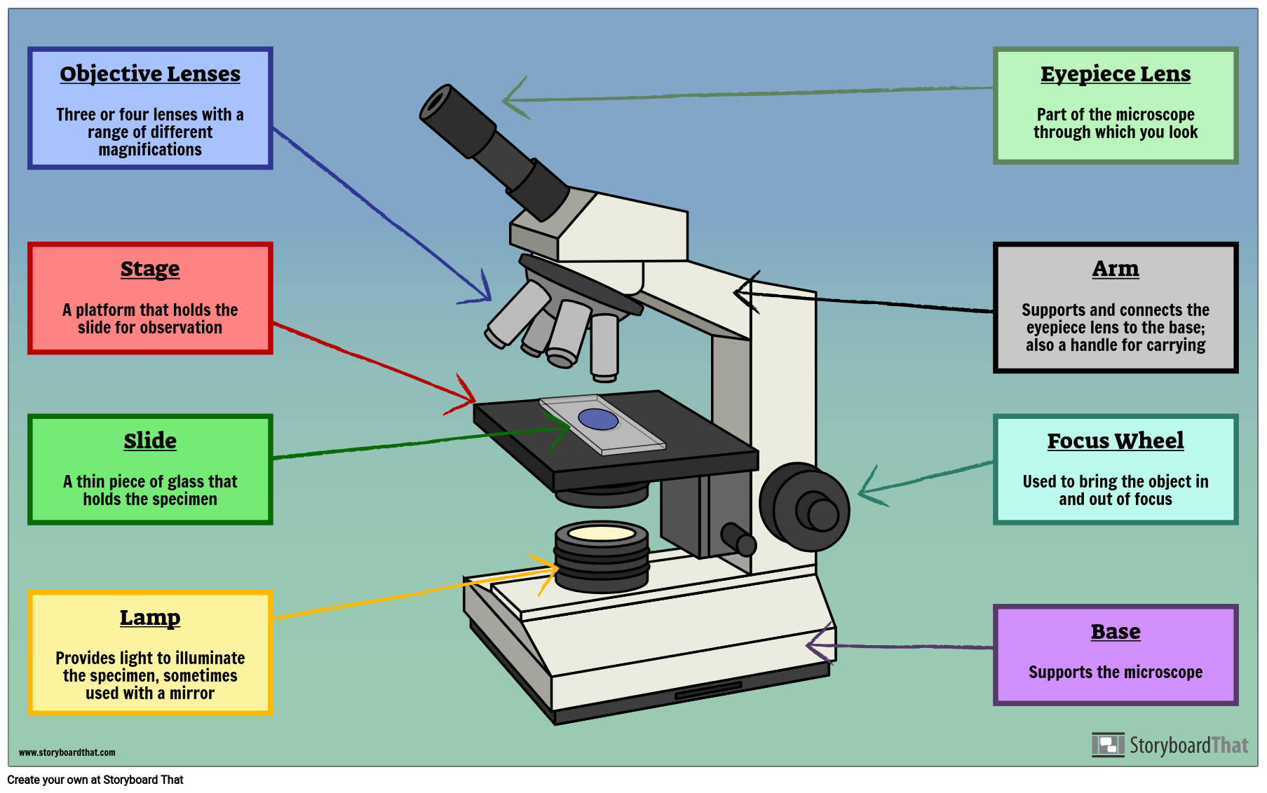
Labelled Microscope with Functions Storyboard by oliversmith
Magnification is a measure of how much larger a microscope (or set of lenses within a microscope) causes an object to appear. For instance, the light microscopes typically used in high schools and colleges magnify up to about 400 times actual size. So, something that was 1 mm wide in real life would be 400 mm wide in the microscope image.
Cells and Microscopes
A Study of the Microscope and its Functions With a Labeled Diagram - Science Struck A Study of the Microscope and its Functions With a Labeled Diagram To better understand the structure and function of a microscope, we need to take a look at the labeled microscope diagrams of the compound and electron microscope.
Types, Parts And Functions Of A Microscope arnoticias.tv
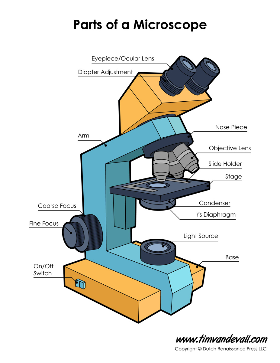
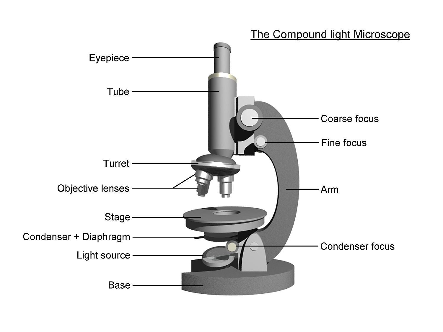
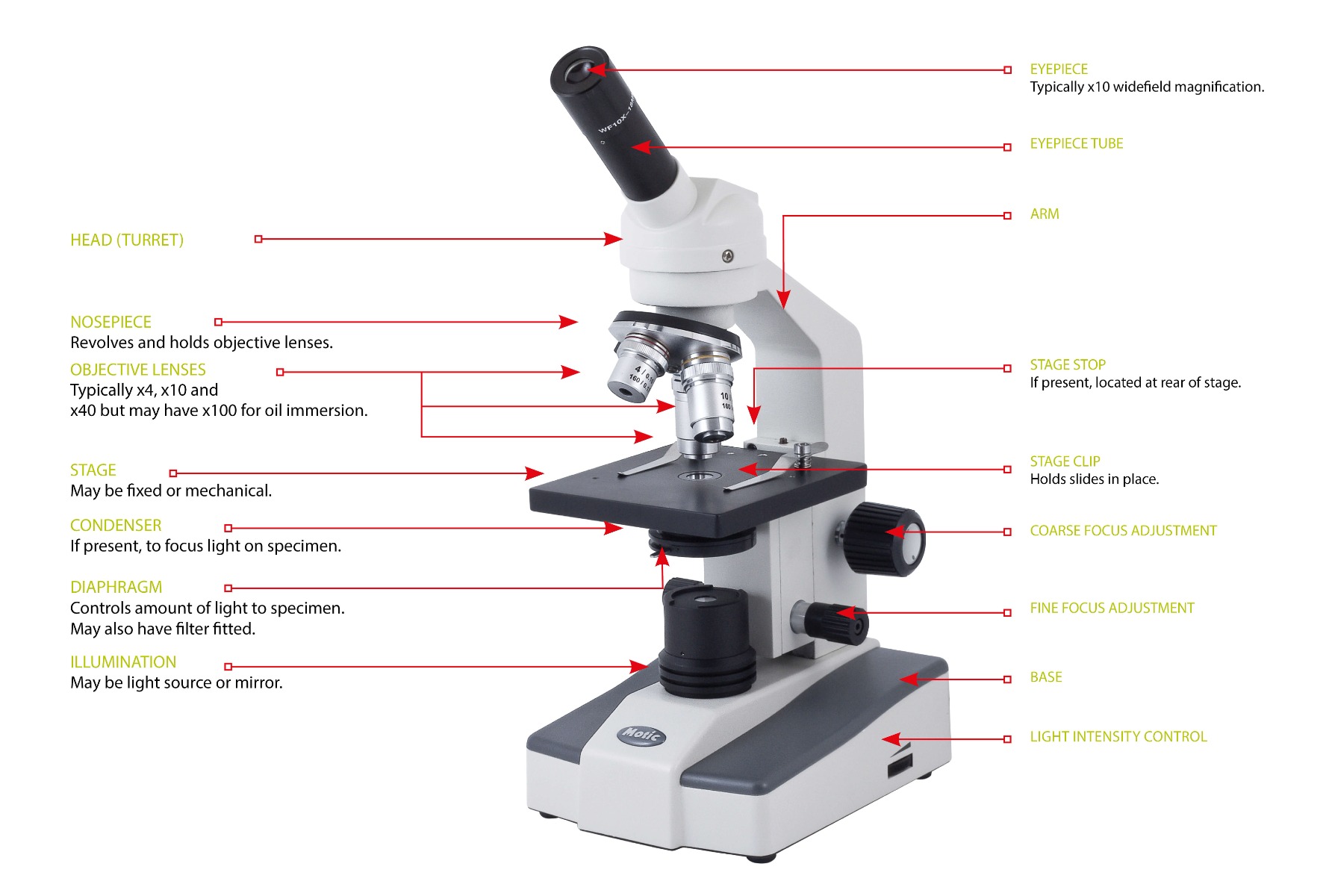
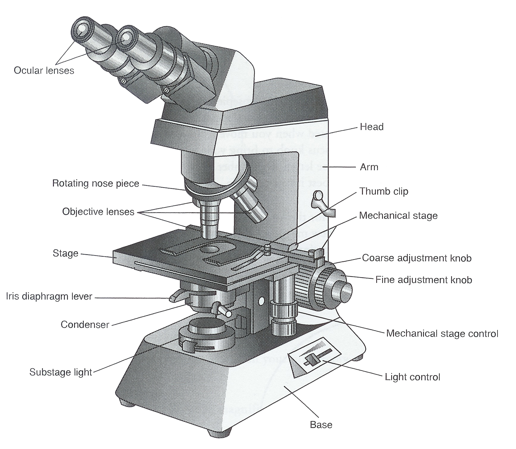
1. Eyepiece 2. Body tube/Head 3. Turret/Nose piece 4. Objective lenses 5. Knobs (fine and coarse) 6. Stage and stage clips 7. Aperture 9. Condenser 10. Condenser focus knob 11. Iris diaphragm 12. Diopter adjustment 13. Arm 14. Specimen/slide 15. Stage control/stage height adjustment 16. On and off switch 17. Base

5 Types of Microscopes with Definitions, Principle, Uses, Labeled Diagrams
The hand magnifying glass can magnify about 3 to 20×. Single-lensed simple microscopes can magnify up to 300×—and are capable of revealing bacteria —while compound microscopes can magnify up to 2,000×. A simple microscope can resolve below 1 micrometre (μm; one millionth of a metre); a compound microscope can resolve down to about 0.2 μm.
Microscope Labelled Diagram Gcse Micropedia Gambaran
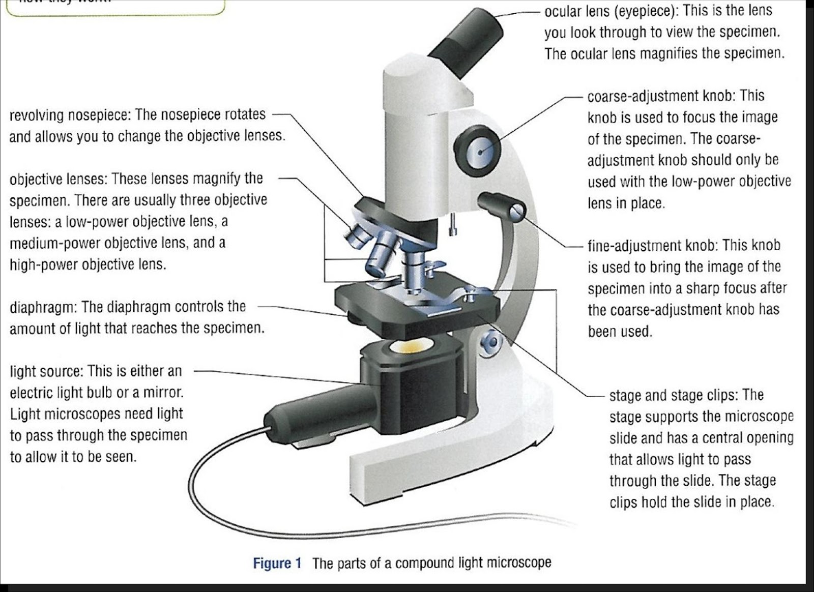
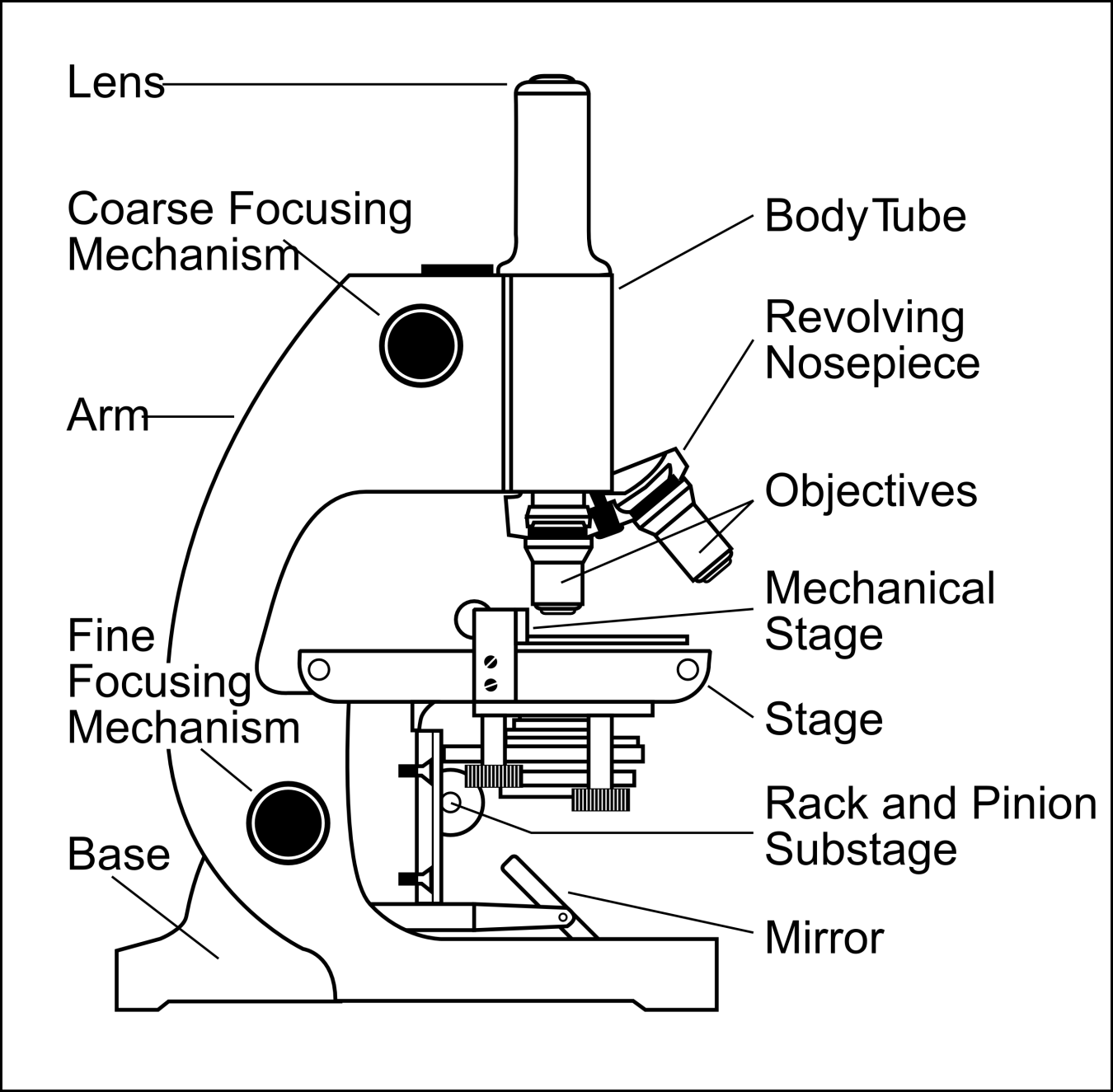
Figure: Diagram of parts of a microscope. There are three structural parts of the microscope i.e. head, arm, and base. Head - The head is a cylindrical metallic tube that holds the eyepiece lens at one end and connects to the nose piece at other end. It is also called a body tube or eyepiece tube.

301 Moved Permanently
See: Labeled Diagram showing differences between compound and simple microscope parts. Structural Components. The three structural components include. 1. Head. This is the upper part of the microscope that houses the optical parts. 2. Arm . This part connects the head with the base and provides stability to the microscope.
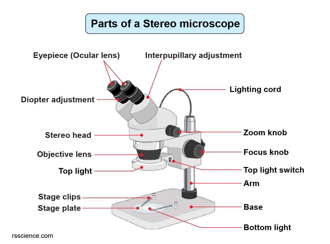
Parts of Stereo Microscope (Dissecting microscope) labeled diagram, functions, and how to use it
The web page titled "Parts of a Microscope with Labeled Diagram and Functions" has the following key takeaways: 🔍 The microscope is an essential tool for scientists, researchers, and medical professionals. 🧬 The main function of a microscope is to provide a magnified view of small objects or organisms, such as bacteria, cells, or tissues.

Parts of a microscope with functions and labeled diagram
a. Mechanical Parts of a Compound Microscope Foot or Base Pillar Arm Stage Inclination Joint Clips Diaphragm Nose piece/Revolving Nosepiece/Turret Body Tube Adjustment Knobs b. Optical Parts of a Compound Microscope Eyepiece lens or Ocular Mirror Objective Lenses

Parts of a Microscope The Comprehensive Guide Microscope and Laboratory Equipment Reviews
Iris diaphragm: Adjusts the amount of light that reaches the specimen. Condenser: Gathers and focuses light from the illuminator onto the specimen being viewed. Base: The base supports the microscope and it's where illuminator is located. How Does a Compound Microscope Work?

The Microscope Its Parts and Their Functions Microscope, Biology lesson plans, Biology lessons
This activity has been designed for use in homes and schools. Each microscope layout (both blank and the version with answers) are available as PDF downloads. You can view a more in-depth review of each part of the microscope here. Download the Label the Parts of the Microscope PDF printable version here.
