Dog anatomy Royalty Free Vector Image VectorStock
Dog Anatomy, Artwork Photograph by Friedrich Saurer Fine Art America
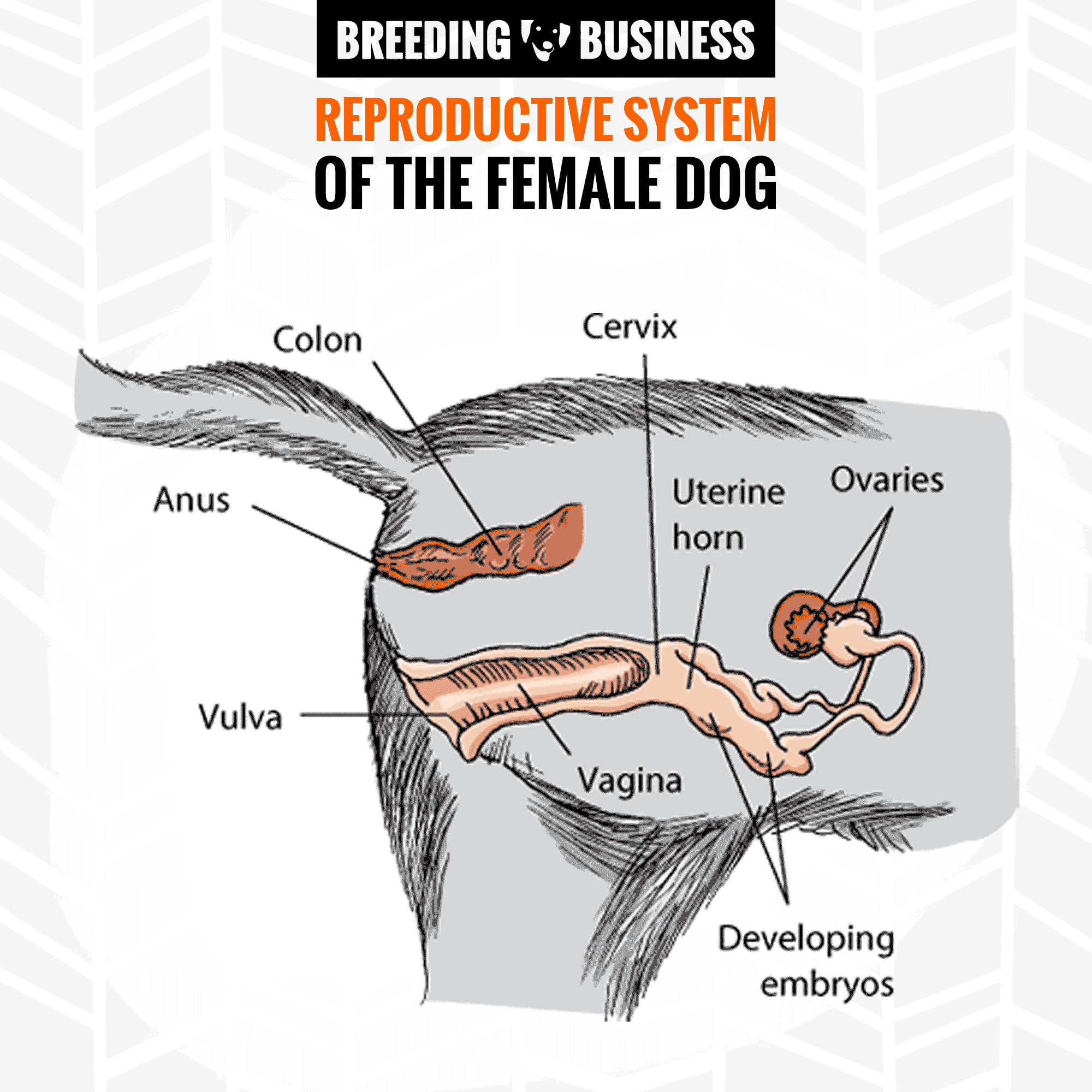
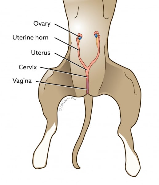
Reproductive system of a female dog The female genital tract includes the vulva, vagina, cervix, uterus, oviducts, and ovaries. The mammary glands, found on the chest and abdomen, are also part of the reproductive system. The oviducts (also called Fallopian tubes) are small tubes that connect the ovaries to the uterus.
Male & Female Dog Reproductive Systems — Organs and Hormones
2021 Ultimate Guide to Dog Anatomy.. urethra and bladder in the excretion of liquid wastes and the reproductive system that includes the female uterus, ovaries, fallopian tubes and vagina and the male testes, epididymis, vas deferens and penis. Lower urinary tract. Female genitalia.
MAJOR MUSCLES IN THE DOG🐕 (origin, insertion&action) Diagram Quizlet
Part 3: Female Genitalia. Abby Brown. Related Learning Objectives. D6.5 Describe and identify the various parts of the female reproductive tract that are found within the abdominal cavity and pelvic region; explain the clinical relevance of these structures. D6.6 Describe the normal parts and placement of the urogenital tract and the.

Dog anatomy Royalty Free Vector Image VectorStock
Dog - Muscles Dog - Thorax/Abdomen/Pelvis Animal - Anatomy atlas: Cardiovascular system Veterinary anatomy - Animal: ANATOMICAL PARTS Abdomen Abdominal aorta Abdominal mammary gland Abdominal mammary region Accessory carpal bone Acromion Adductor muscle Ala of ilium; Wing of ilium Ala of nose Anconeus muscle Antebrachial region Aortic arch
Bladder Health Herbsmith
Anatomy of the Dog Vagina. The outer portion of a female dog's reproductive tract is called the vulva. It consists of two labia (thick folds of tissue) that are connected at the top and bottom. The vestibule lies just inside of the vulvar opening. The vagina opens into the vestibule, as does the urethra—the tube that drains the bladder.
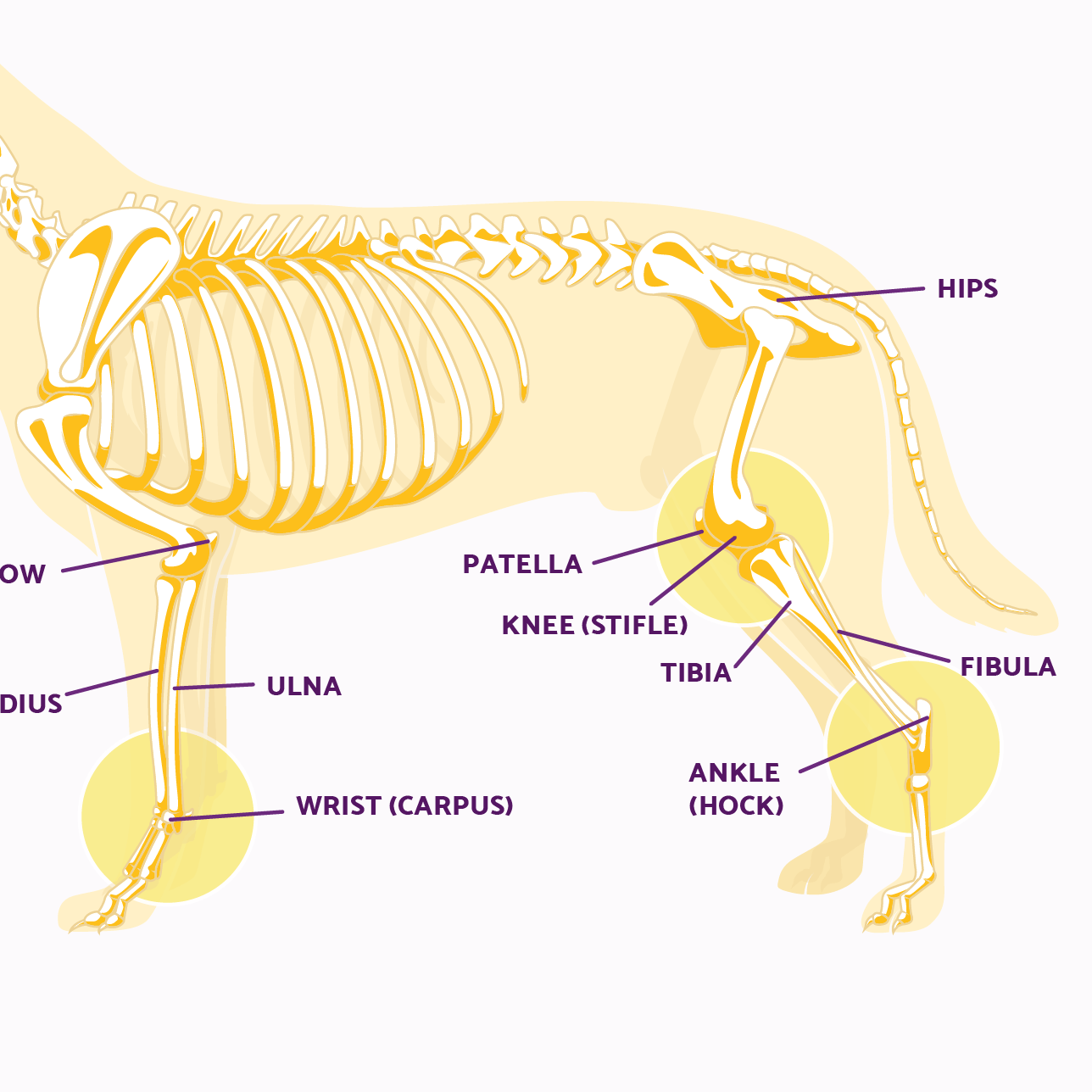
Dog Leg Anatomy in Human Speak Ortho Dog
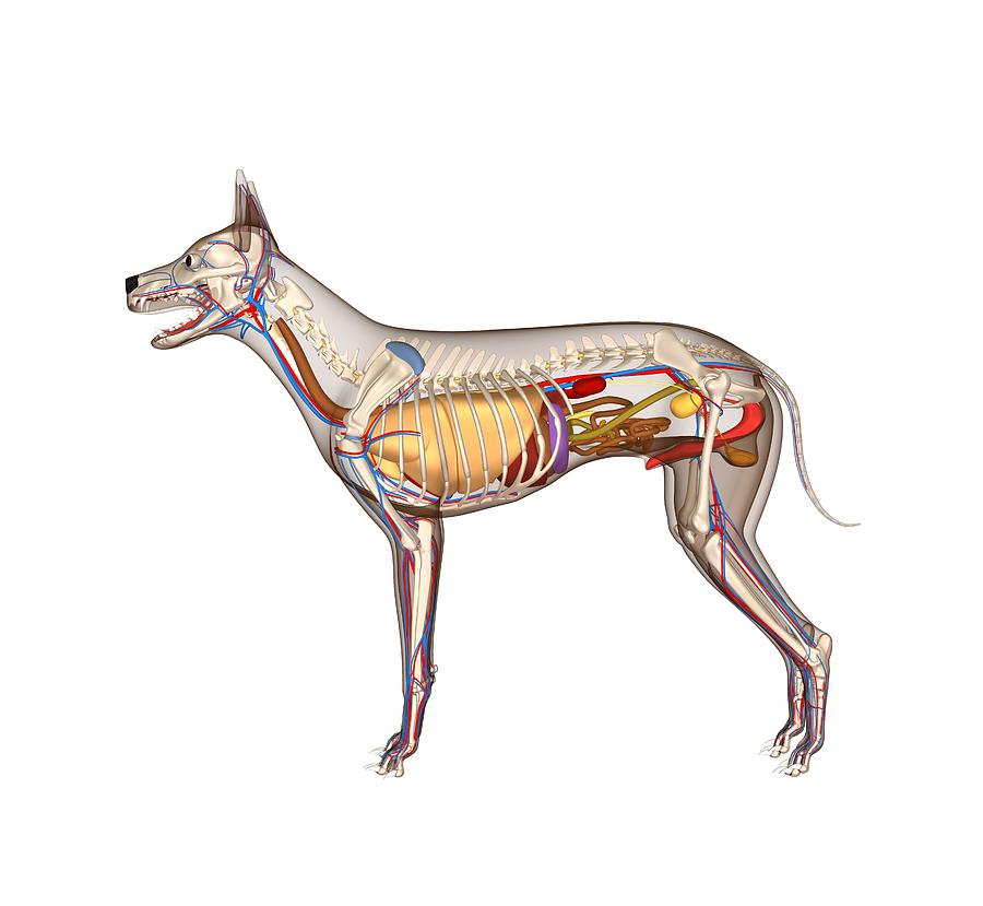
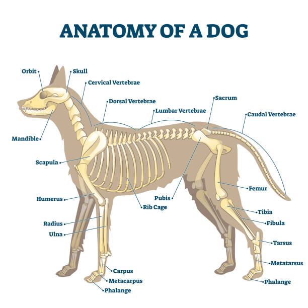
Quick idea: in this article, you will learn the location of different organs from the different systems (like skeletal, digestive, respiratory, urinary, cardiovascular, endocrine, nervous, and special sense) of a dog with their important anatomical features.
4,100+ Dog Anatomy Stock Photos, Pictures & RoyaltyFree Images iStock
Intersex Genitalia - Posterior View. The external genitalia indicate either a megaclitoris or hypoplasia of the penis. An os penis or os clitoris may be palpable. A catheter has been placed in the urethra. The dorsal commissure of the vulva is pulled up and the labiae are spread laterally with forceps. Shille VM (1980)
Spaying in Dogs VCA Animal Hospital
Anatomy and physiology of the female dog The vaginal vestibule, a short entryway into the vagina, is oriented at a 60-degree angle to the horizontal (upward, toward the spine, and forward, toward the head). Thus, to pass a speculum or catheter into the vagina requires that it be initially oriented at this upward angle, and if a female needs.
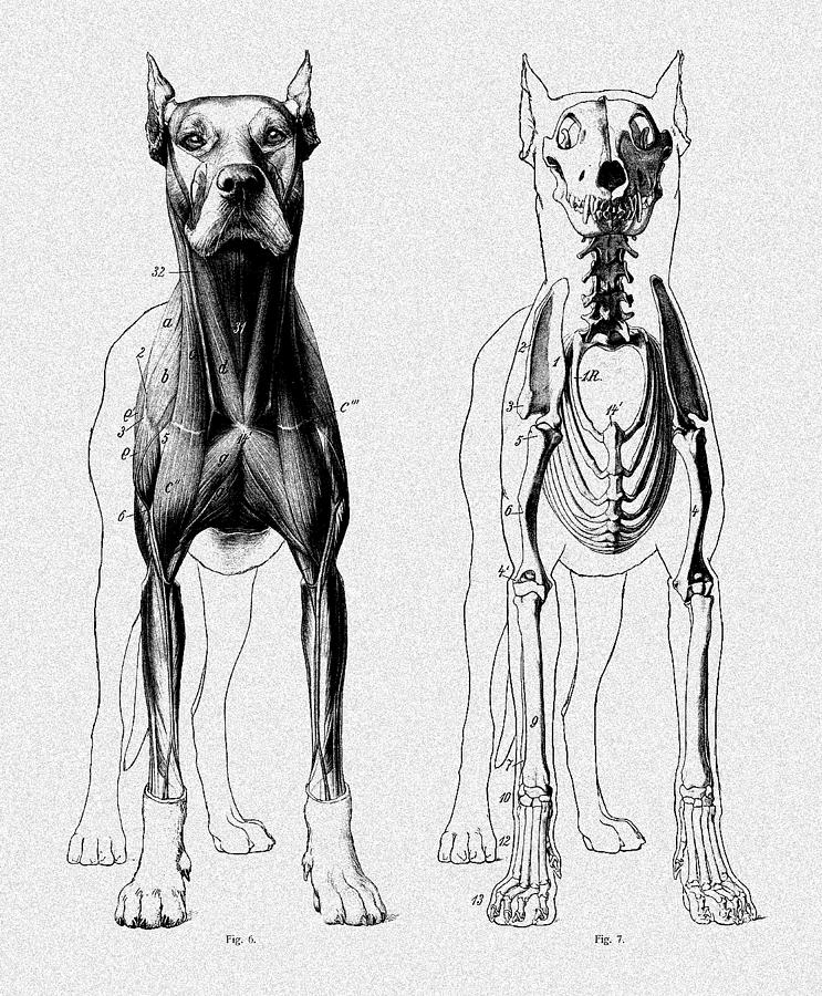
Vintage Dog Anatomy Scientific Illustration Digital Art by Sandra McGinley Fine Art America
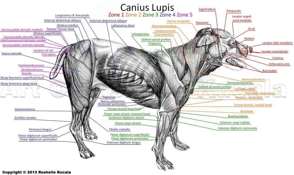
Whereas giant breeds can take between 18 months and 2 years for their growth plates to fuse. Speaking of skeletons, a dog has 320 bones in their body (depending on the length of their tail) and around 700 muscles. Muscles attach to bones via tendons. Depending on the breed of dog, they will have different types of muscle fibers.
Dog Domestication Britannica
The male dog dismounts the female at this time. The dogs can remain in this position from a few minutes to an hour, and it is recommended not to try and separate them as it can cause injury to their organs. A dog's anatomy is not very different from any other mammal's.
DOG ANATOMY 3D model by zorrenhimself [a79b90c] Sketchfab
The vulva in a female dog is located just beneath the anus and above the urethral opening. It can be visualized as a small, slit-like structure positioned in the lower part of the dog's abdomen. A diagram can provide a helpful visual representation of its exact location. Understanding the Anatomy: Exploring the Vulva on a Female Dog

Dog Anatomy Diagram Quizlet
The female dog anatomy external organ is the vulva, which opens to the vagina. A pregnant female dog's anatomy includes two ovaries, which produce eggs, the cervix, fallopian tubes, and the uterus. The uterus becomes the womb for her puppies during their gestation period. Dorling Kindersley via Getty Images
Male & Female Dog Reproductive Systems — Organs and Hormones
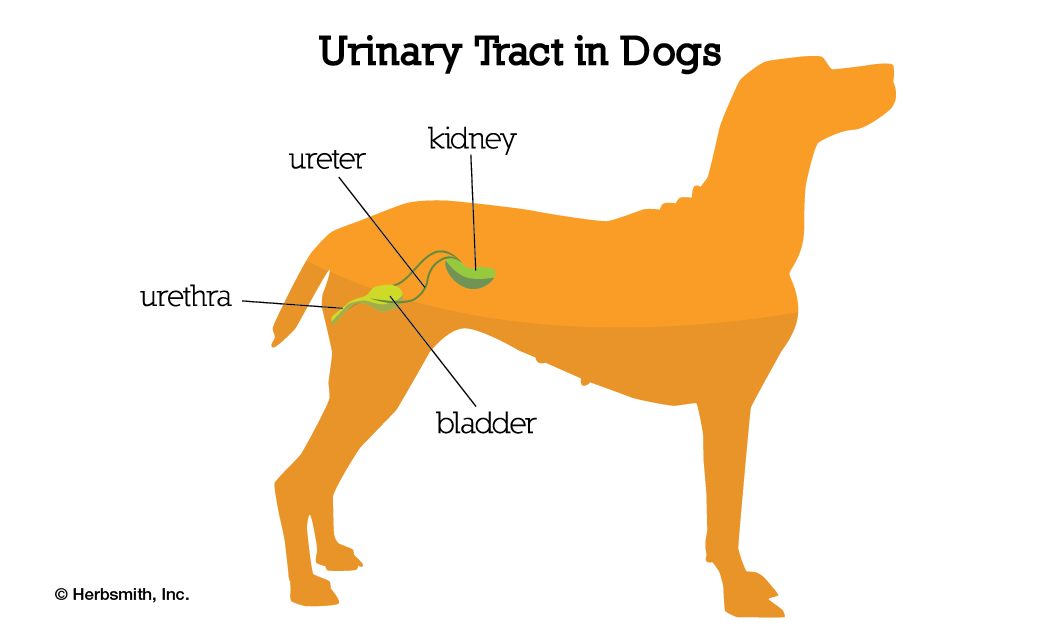
Urinary system in female dogs The urinary system or tract includes the kidneys, the ureters (tubes that connect the kidneys to the bladder), the bladder, and the urethra (the tube through which urine exits the body). The urinary system has several important functions.

Pin en Pet Remedies
In this video, we are going to open the abdominal cavity and describe the female Reproductive System of the dog which consists of ovaries, uterine tubes, ute.
4,100+ Dog Anatomy Stock Photos, Pictures & RoyaltyFree Images iStock
Here are some key points to help you understand the female dog anatomy and the estrous cycle: Ovaries: Female dogs have two ovaries, which are responsible for producing eggs and releasing hormones involved in the estrous cycle. Uterus: The uterus is where the fertilized eggs develop into puppies during pregnancy. It undergoes changes during.

Best Dog Anatomy Stock Photos, Pictures & RoyaltyFree Images iStock
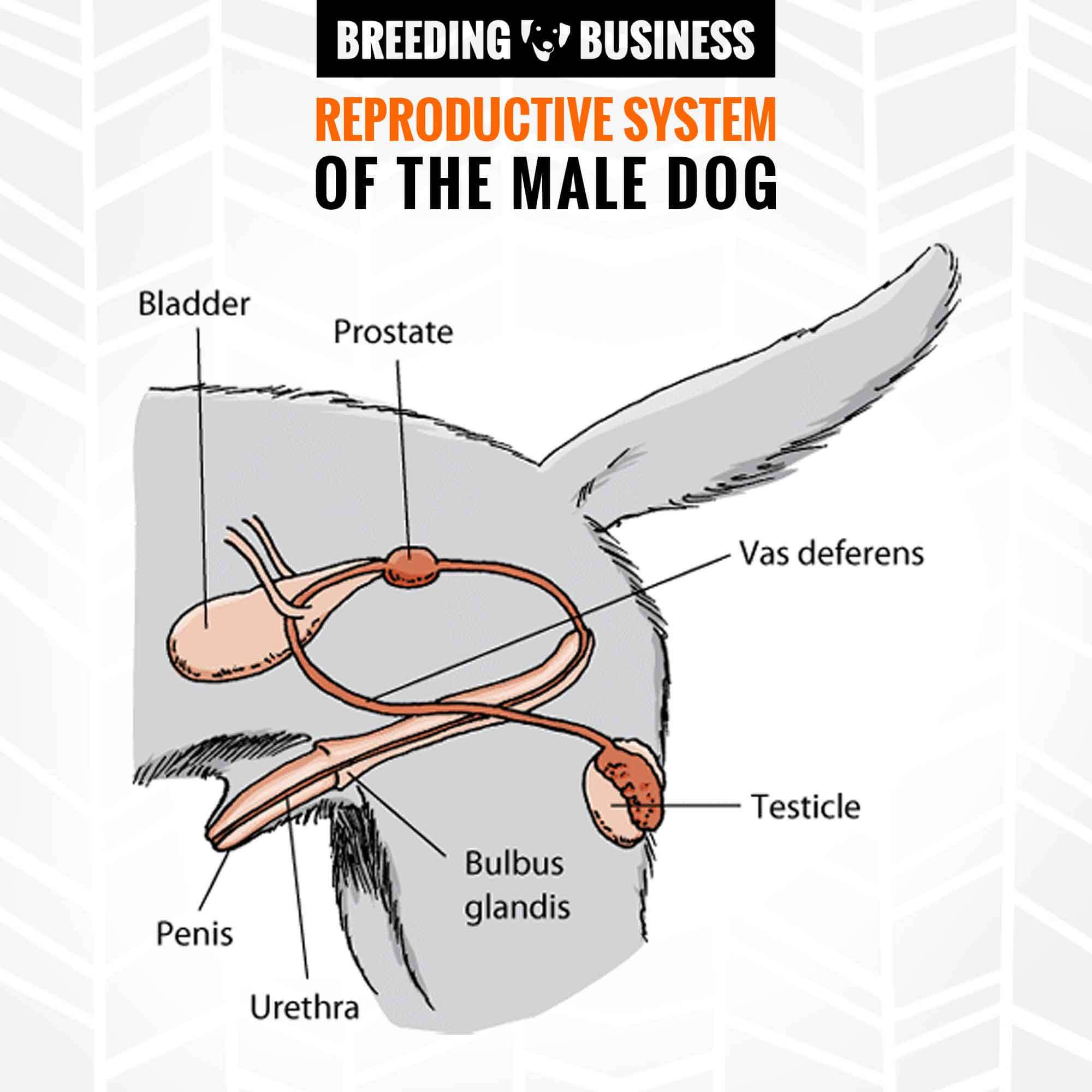
This gland produces prostatic fluid, which feeds the sperm and helps maintain them in their passage into the female dog. The male dog urethra therefore carries sperm from the testicles into the penis. The urethra also carries urine from the bladder to the penis.
