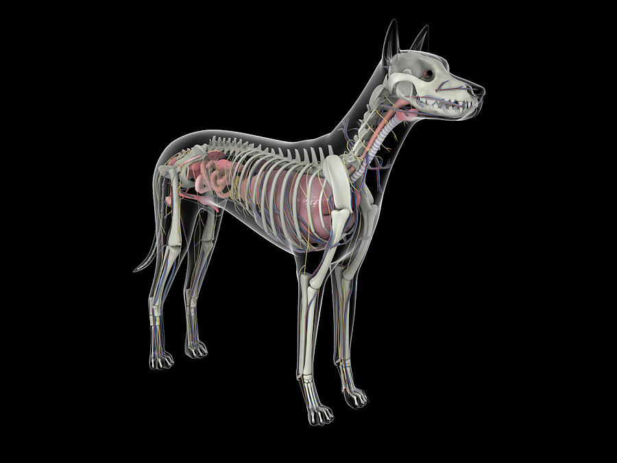
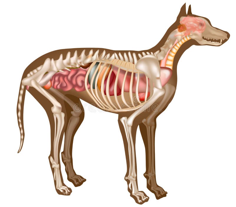
Anatomy of a male dog crosssection, showing the skeleton and internal

Глубокие мышцы, внутренние органы собаки Dog Muscles & Internal
Whereas giant breeds can take between 18 months and 2 years for their growth plates to fuse. Speaking of skeletons, a dog has 320 bones in their body (depending on the length of their tail) and around 700 muscles. Muscles attach to bones via tendons. Depending on the breed of dog, they will have different types of muscle fibers.
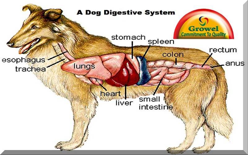
How is a Dog Digestive System Functioning? Growel Agrovet Private Limited
The internal anatomy of dogs, is very similar to the anatomy of other carnivorous mammals such as the cat. Dogs have a well-developed brain which is composed of different parts. The cerebrum is the part of the dog's brain which performs functions such as learning.
Dog Digestive Process and what the stages are and how it works
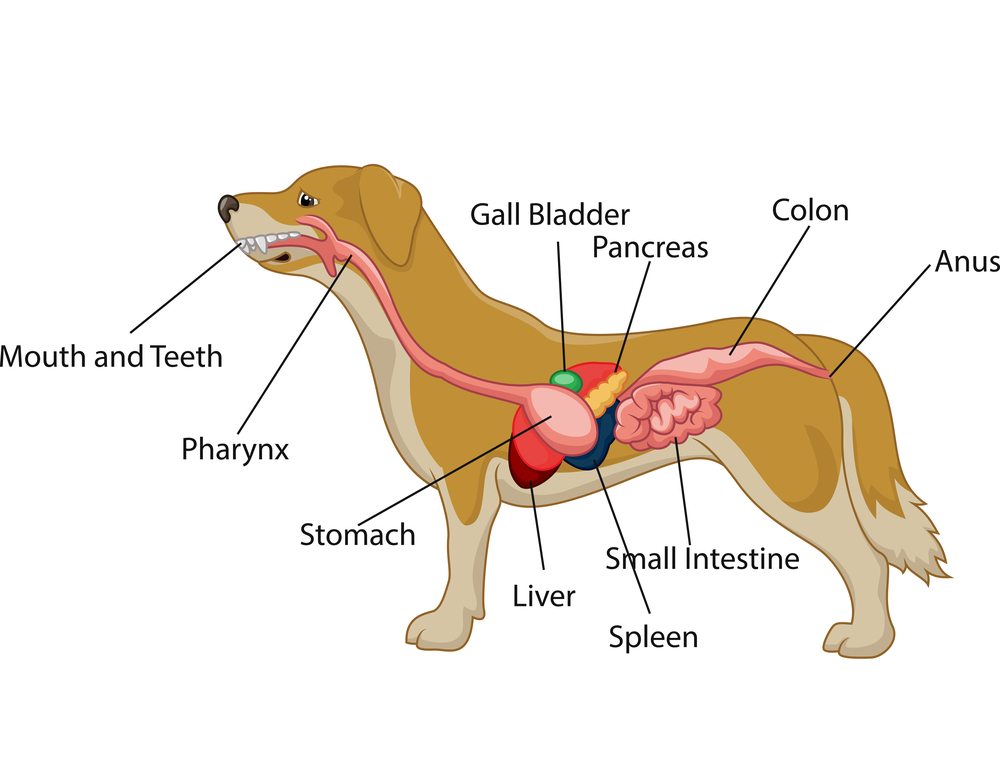
The internal organs of a dog include the heart, lungs, liver, kidneys, stomach, intestines, and reproductive organs. These organs work together to keep the dog healthy and functioning properly. For example, the heart pumps blood throughout the body, while the kidneys filter waste products from the blood.
Dog Anatomy With Internal Organs Photograph by Stocktrek Images Fine
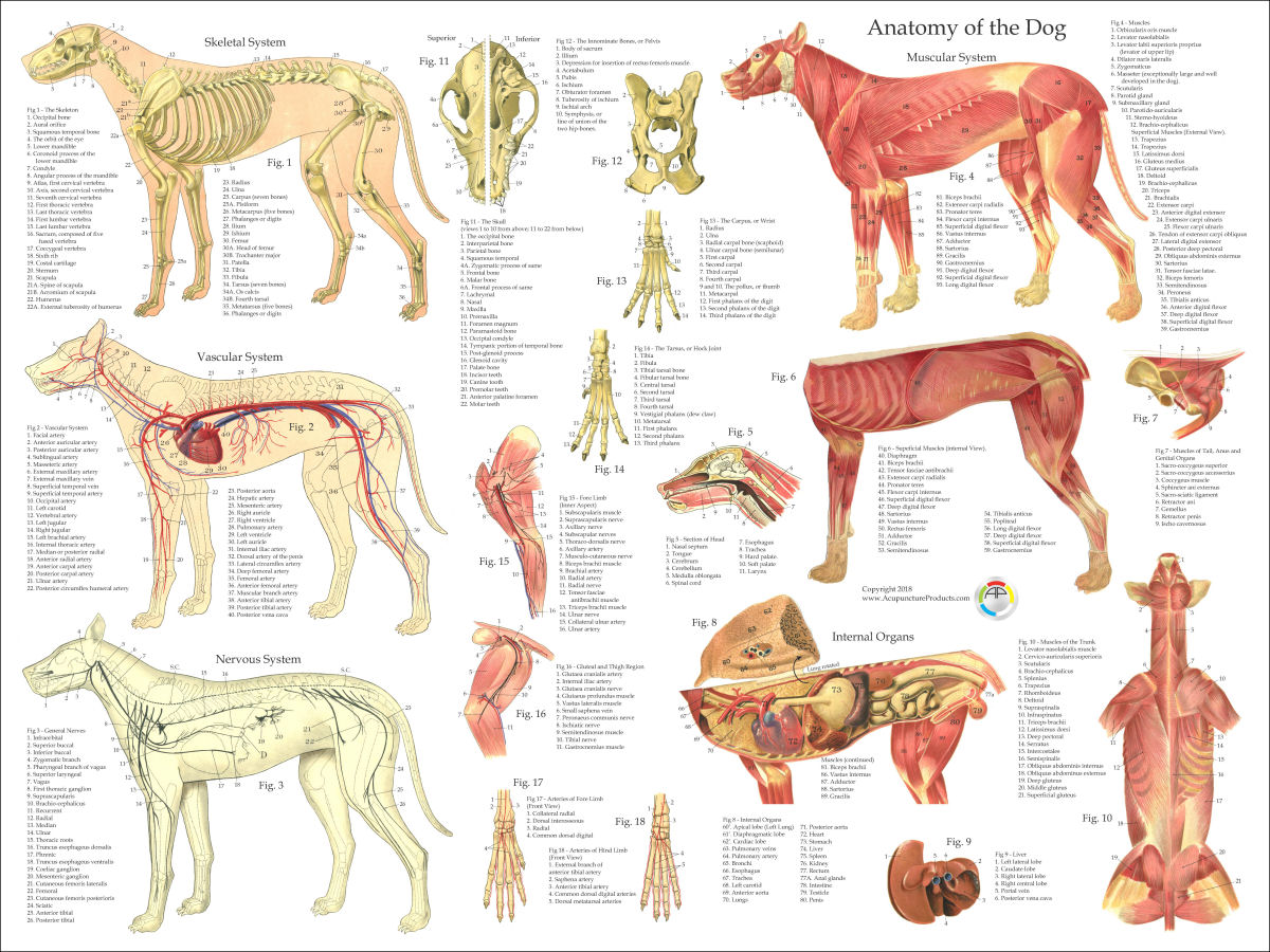
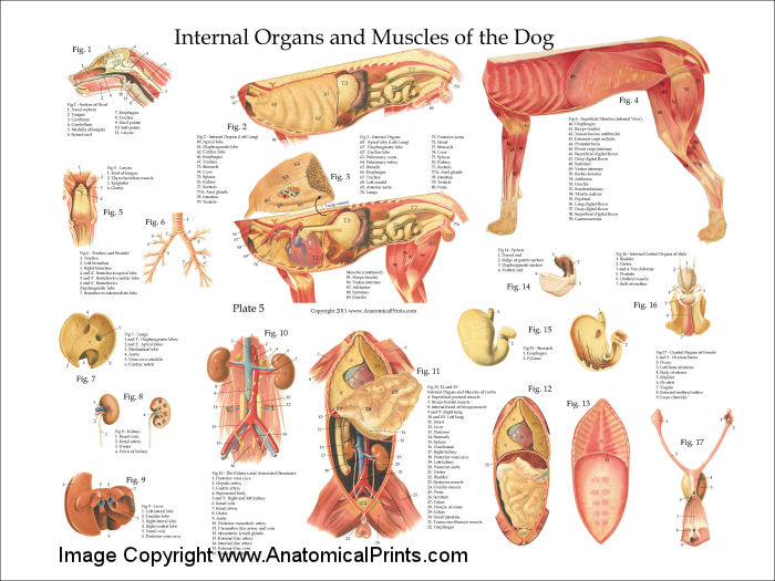
This veterinary anatomy module contains 608 illustrations on the canine myology. Here are presented scientific illustrations of the canine muscles and skeleton from different anatomical standard views (lateral, medial, cranial, caudal, dorsal, ventral / palmar.). Some fascias, tendons, ligaments, joints were labeled.

A4 Veterinary Poster u00 Internal Organs Of The Dog (Animal Anatomy
Dog anatomy comprises the anatomical studies of the visible parts of the body of a domestic dog.Details of structures vary tremendously from breed to breed, more than in any other animal species, wild or domesticated, as dogs are highly variable in height and weight. The smallest known adult dog was a Yorkshire Terrier that stood only 6.3 cm (2.5 in) at the shoulder, 9.5 cm (3.7 in) in length.

Pin em Anatomy
The canine tibia is the major bone in the crus. The triangular proximal tibia is wider than the distal cylindrical tibia. Medial and lateral tibial condyles, an intercondylar eminence, and a tibial tuberosity are on the proximal tibia. The tibial plateau slopes distally from cranial to caudal.

Dog Anatomy Skeleton Animaltia
The anatomy of the temporal bone and the ear is complex as this region concentrates a large number of bony, muscular, articular, vascular and nervous structures. The purpose of the current anatomy module is to describe the normal anatomy of the inner and middle ear of the dog as depicted using CT of the temporal bone. Material and methods
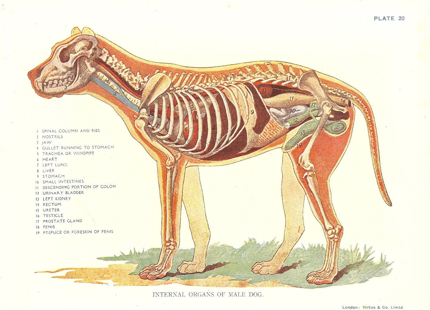
Dog Veterinary Print 1920s Internal Organs Of Male Dog
2021 Ultimate Guide to Dog Anatomy. As the pace of veterinary advancement accelerates, even the most experienced veterinary teams are challenged to keep up with all the changes that impact their practice. Veterinary teams need practical, concise and relevant visual aids at their fingertips while in practice, helping them to prescribe the right.
Canine Internal Anatomy Chart. Anatomy of Dog with Inside Organ
A dog's physical anatomy is designed to help them navigate their environment and perform various tasks. Their bodies are made up of many different parts, including their skeleton, muscles and internal organs. One of the most important parts of a dog's anatomy is their skeleton.

Anatomy of a male dog crosssection, showing the skeleton and internal
The spleen is another clinically important organ in dog internal anatomy. There is a roughly human foot-print-shaped structure spleen present in a dog. The ventral end is wider than the dorsal end of the dog's spleen. Again, the dog's spleen location is variable except for the upper end, which is below the proximal end of the last rib.
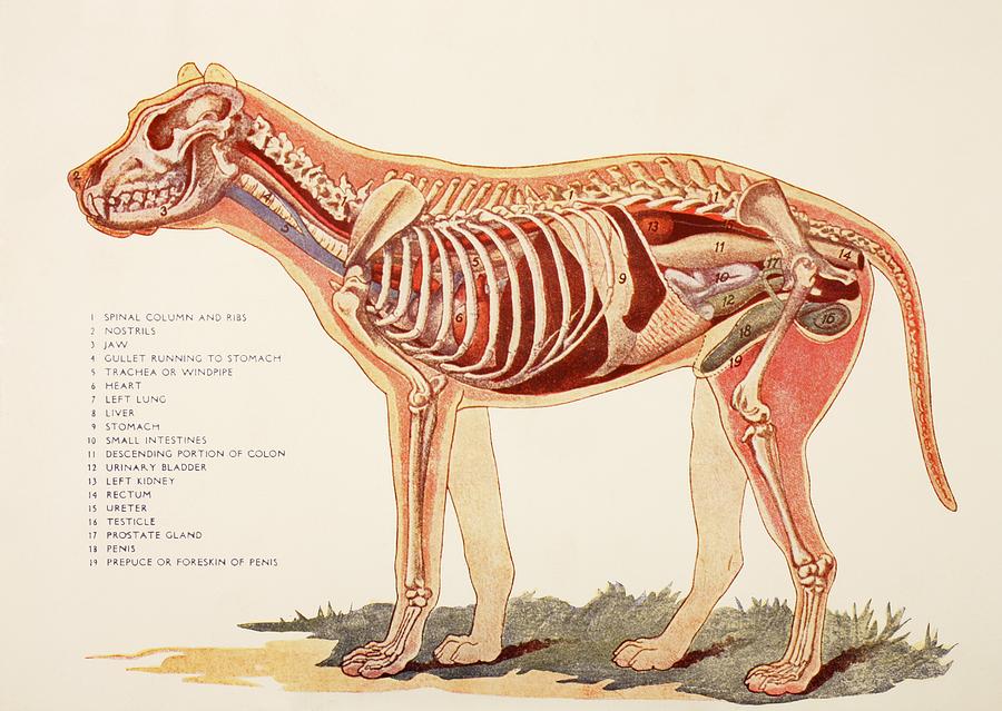
Internal Organs Of A Male Dog. From Photograph by Ken Welsh Pixels
Xiphoid region (Cranial abdominal region) Zygomatic bone. Zygomatic gland. Zygomatic region. Radiographic anatomy: labeled images in the transverse plane of a healthy dog's whole body, using tomodensitometry. Introduction to the anatomy of the skull, thorax, abdomen, pelvic cavity, muscles and blood vessels: main anatomical structures identified.
Dog Anatomy Poster
It provides information about a dog's skeletal, reproductive, internal, and external anatomy, along with accompanying labeled diagrams. After mating, dogs experience something called a copulatory tie, wherein they remain in the coital position. The male dog dismounts the female at this time. The dogs can remain in this position from a few.
Dog Internal Anatomy Poster
Anatomy atlas of the canine general anatomy: fully labeled illustrations and diagrams of the dog (skeleton, bones, muscles, joints, viscera, respiratory system, cardiovascular system). Positional and directional terms, general terminology and anatomical orientation are also illustrated.

Pin en Pet Remedies
In addition to the world's most segmented dog anatomy, the Table Vet also includes a diverse library of animal cases.. The Anatomage Dog is the first highly detailed dog anatomy atlas that comprehensively features internal organs, including vascular systems and muscular-skeletal structures. Originating from real dog data, the Anatomage Dog.
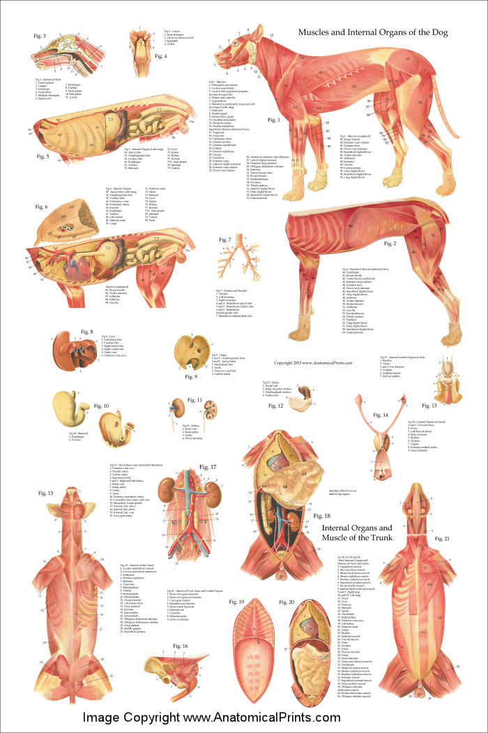
Dog Internal Anatomy Poster 24 x 36
Anatomy of internal dog throat. In this section, you will learn the anatomical facts of the different organs and structures of the internal dog throat. First, I will start with the other cartilages of the dog's larynx. Then I will describe the anatomical facts of the trachea and esophagus with the diagrams.

dog anatomy Dog Care Training Grooming
A female dog's reproductive system has similar organs as a human's. The female dog anatomy external organ is the vulva, which opens to the vagina. A pregnant female dog's anatomy includes two ovaries, which produce eggs, the cervix, fallopian tubes, and the uterus. The uterus becomes the womb for her puppies during their gestation period.
